Lung Hilar Anatomy
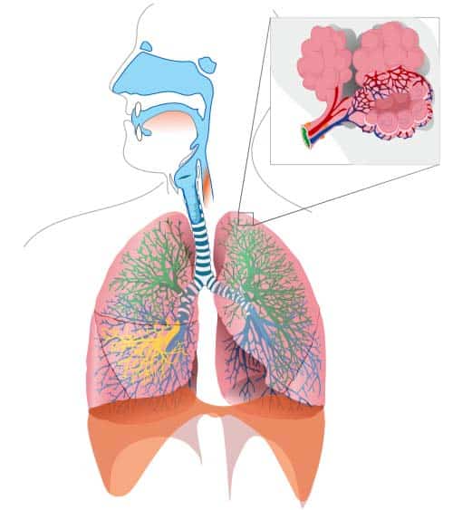
Each lung consists of. The resulting system of tubules resembles an inverted tree.
Untitled
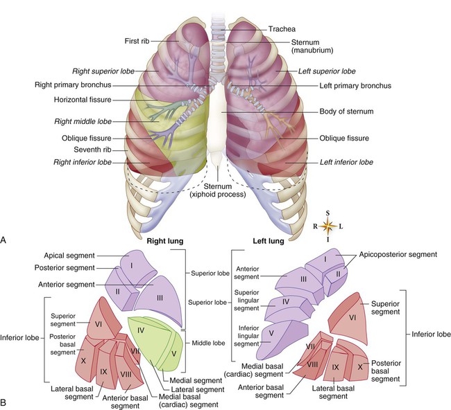
Lobes two or three these are separated by fissures within the lung.
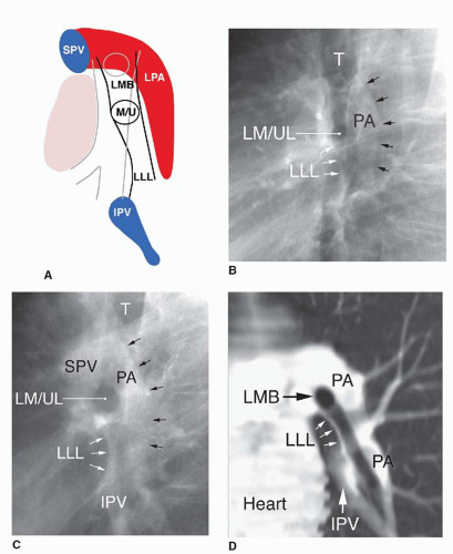
Lung hilar anatomy. Below this is the left main bronchus. The hilar region of. Tests to evaluate the hilum.
Base the inferior surface of the lung which sits on the diaphragm. Lung roots lie opposite to t5 t7 vertebrae. Apex the blunt superior end of the lung.
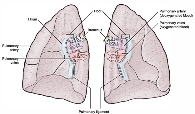
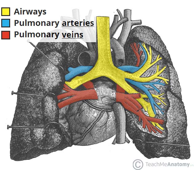
The lungs are pyramid shaped paired organs that are connected to the trachea by the right and left bronchi. The structures of the lung root are embedded in the connective tissue and surrounded by extension. The lung hila or roots are found on the medial aspect of each lung.
The left and right lung roots are similar but not identical. Hilum of the lung. Anatomy and abnormalities anatomy of the hilum.
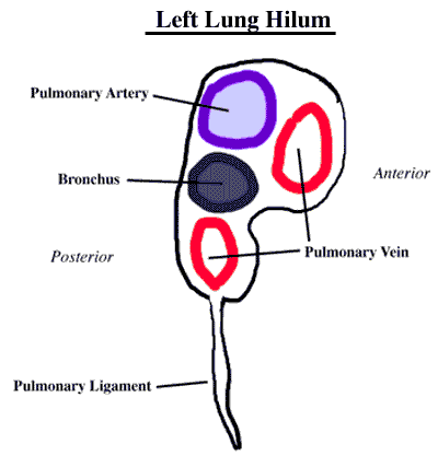
The hila are not symmetrical but contain the same basic structures on each side. In the left hilum the left pulmonary artery occupies the upper part. Hilum of lung a triangular depression where the structures which form the root of the lung enter and leave the viscus hilum of lymph node the portion of a lymph node where the efferent vessels exit.
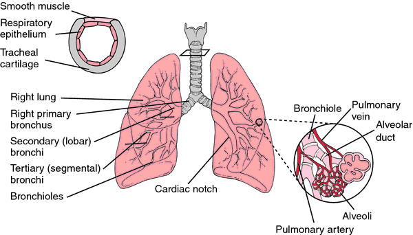
The root of the lung is located at the hilum of each lung just above the middle of the mediastinal surface and behind the cardiac impression of the lung. These structures pass through the narrow hila on each side and then branch as they widen out into the lungs. In lung to its apex is the hilum the point at which the bronchi pulmonary arteries and veins lymphatic vessels and nerves enter the lung.
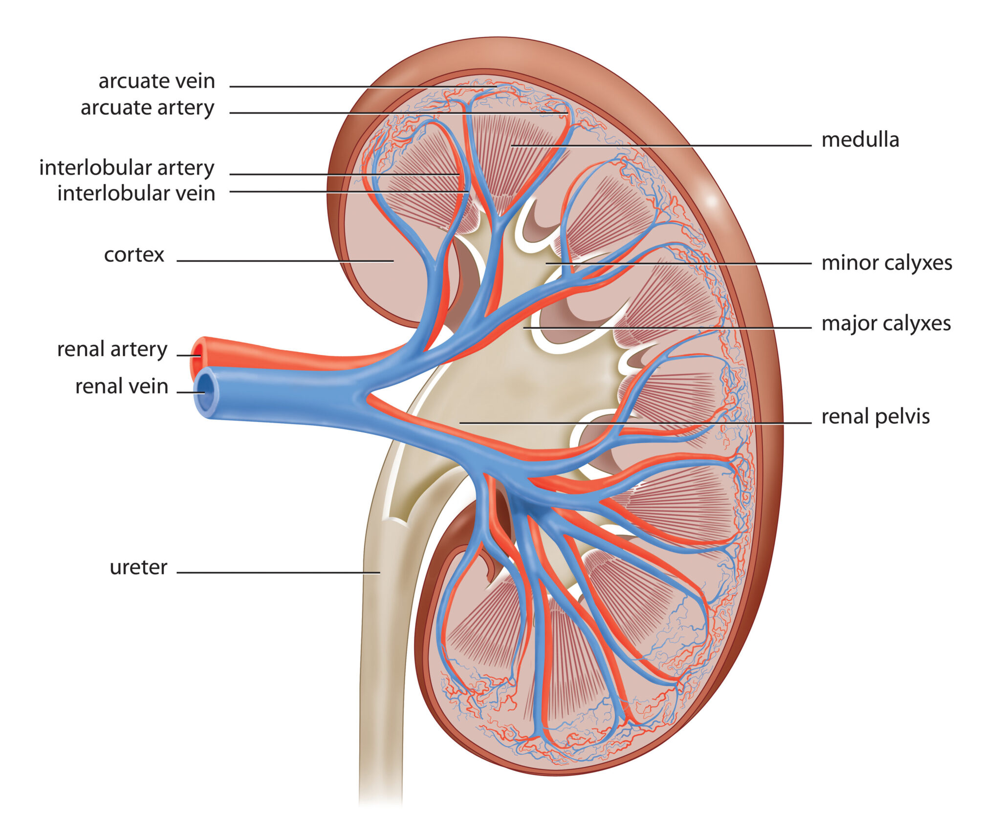
Abnormalities in the hilum are usually noted on imaging. The diameters of the bronchi diminish eventually to. The rib cage is separated from the lung by a two layered membranous coating the pleura.
The hila lung roots are complicated structures mainly consisting of the major bronchi and the pulmonary veins and arteries. Describe the root and hilum of lungs. It is nearer to the back than the front.
The root of the lung is connected by the structures that form it to the heart and the trachea. The diaphragm is the flat dome shaped muscle located at the base of the lungs and thoracic cavity. Surfaces three these correspond to the area of the thorax that they face.
On the inferior surface the lungs are bordered by the diaphragm. The main bronchus subdivides many times after entering the lung. The hilum is the large triangular depression where the connection between the parietal pleura and the visceral pleura is.
Lung root consists of the structures passing to and from the hilum of the lung to the mediastinum. Both the right and the left lung have a hilum which lies roughly midway down. Gross anatomy of the lungs.
Gross anatomy left hilum.
 Pulmonary Anatomy And Physiology Nurse Key


 Anatomy Atlas Netter Docsity
Hilum Of Lung
 Root Of The Lung Wikipedia
Summary Netter S Anatomy Lecture Lungs Dbs 8110 Studocu
 Vats Right Upper Lobe Rul Segmentectomy Master
 Pulmonary Artery Wikipedia
 Ecr 2015 C 2530 Reminding The Pulmonary Hila From
 The Lungs Position Structure Teachmeanatomy
 Lung Hilus Definition Of Lung Hilus By Medical Dictionary
 The Pulmonary Hila Radiology Key
 Hilum Anatomy Britannica
 Pulmonary Vascular System And Pulmonary Hilum Sciencedirect
 Easy Notes On Lungs Learn In Just 4 Minutes Earth S Lab
 Pulmonary Vascular System And Pulmonary Hilum Sciencedirect
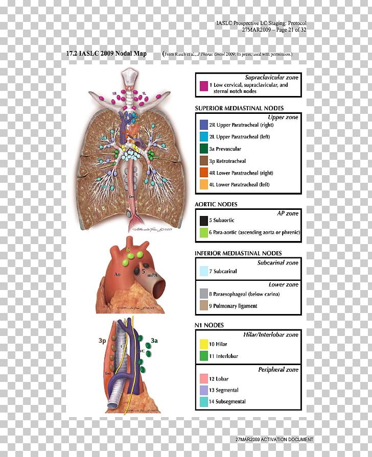 Lymph Node Lung Cancer Staging Lung Cancer Staging Anatomy

What Does Bilateral Hilar Congestion In A Chest X Ray
 Dissector Answers Superior Mediastinum Lungs
 The Lungs Position Structure Teachmeanatomy

Belum ada Komentar untuk "Lung Hilar Anatomy"
Posting Komentar