Shoulder Anatomy Posterior
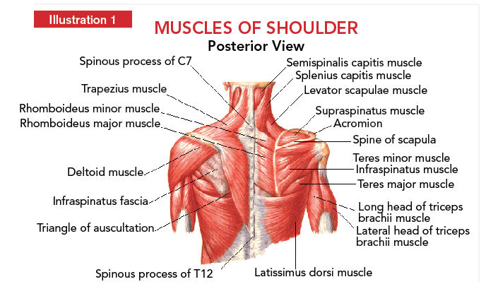
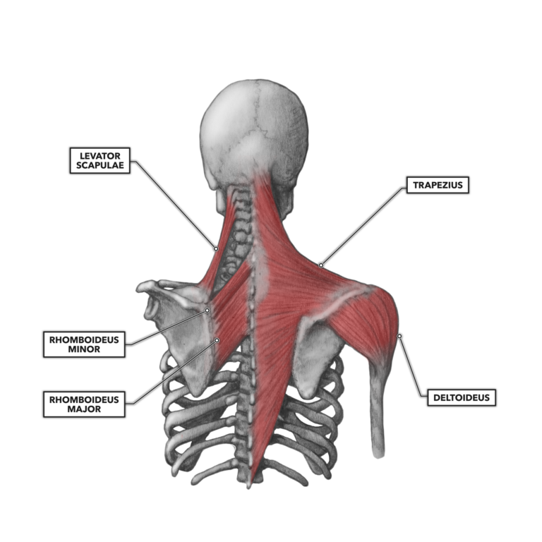
Boney structures shown are the acromion of the scapula the clavicle and the humerus. Pulls raises and adducts the scapula.
 3 Uniquely Powerful Exercises To Address The Root Cause Of
Nerves and blood vessels supply the muscles and bones of the shoulder.
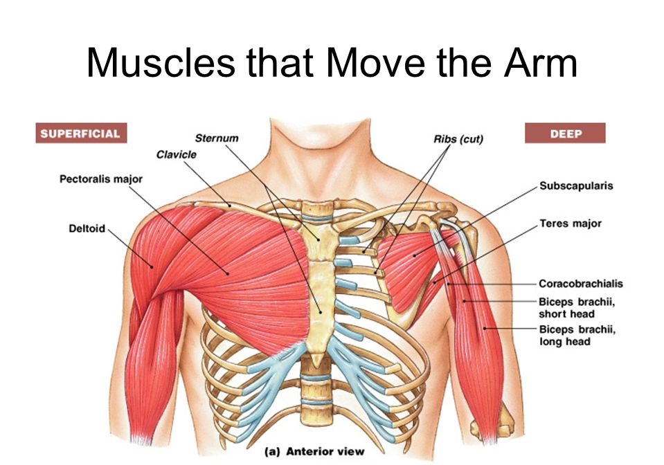
Shoulder anatomy posterior. The shoulder is the group of structures in the region of the joint. On the anterior side of the shoulder the coracobrachialis serratus anterior pectoralis major and pectoralis minor muscles work as a group to flex and adduct the scapula and humerus anteriorly toward the sternum. Rotates the scapula raises the arm.
Trapezius rotates the scapula raises the arm. Other important bones in the shoulder include. Pulls rhomboideus major raises and adducts the scapula.
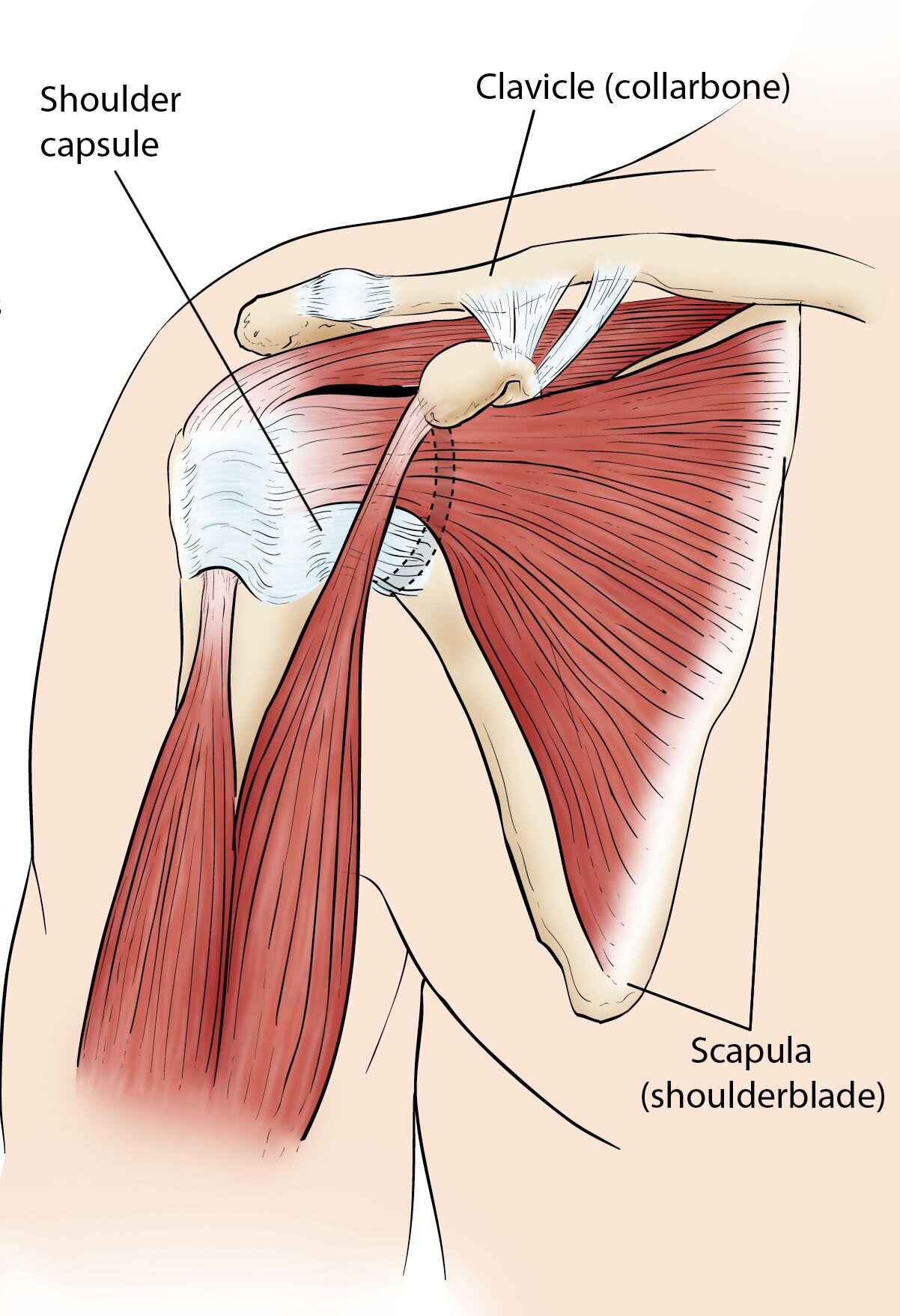
The shoulder joint is formed where the humerus upper arm bone fits into the scapula shoulder blade like a ball and socket. Acute posterior shoulder dislocations are less common than anterior dislocations but more commonly missed 50 of traumatic posterior dislocations seen in the emergency department are undiagnosed. The shoulder complex is composed of many different tissue types and it is the connective tissue that provides the supportive framework for the shoulders many functions.
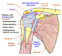
The acromion is a bony projection off the scapula. The clavicle collarbone meets the acromion in the acromioclavicular joint. There is a dissection assistance pdf file that you can use to assist you in your lab preparation.
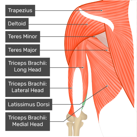
Epidemiology incidence 2 to 5 of all unstable shoulders. Adducts and rotates the arm medially. The latissimus dorsi and teres major on the posterior side extend and adduct the arm towards the vertebrae of the back.
Posterior shoulder instability dislocation. Posterior shoulder the following video will walk you through the six steps to dissecting the posterior shoulder. In human anatomy the shoulder joint comprises the part of the body where the humerus attaches to the scapula the head sitting in the glenoid cavity.
Nerves carry signals from the brain to the muscles to move the shoulder and carries signals from the muscles back to the brain about pain pressure and temperature. The different types of connective tissues in the shoulder are bone articular cartilage ligaments joint capsules and bursa see gross anatomy. The shoulder joint ligaments shown are the acromioclavicular ligament coracoclavicular ligament the superior transverse scapular ligament and the joint capsule or glenohumeral ligaments.
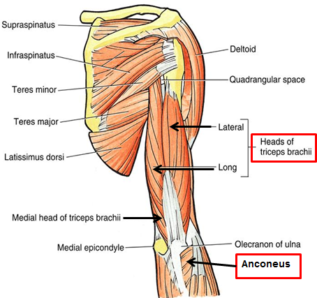
 Triceps Brachii Muscle Medial Head
 The Buford Complex
 Muscle Diagrams Division Of Natural Sciences
 Posterior Shoulder Anatomy Diagram Shoulder Muscle Anatomy
Methods For Manual And Self Stretching Of The Posterior
 Shoulder Muscles Attachment Nerve Supply Action
 Shoulder Development In Strength Training Coach Athletic
 Shoulder Wikipedia
 Shoulder Joint Ligaments Posterior Medical Art Library
 Posterior View Of The Shoulder Shoulder Anatomy Shoulder
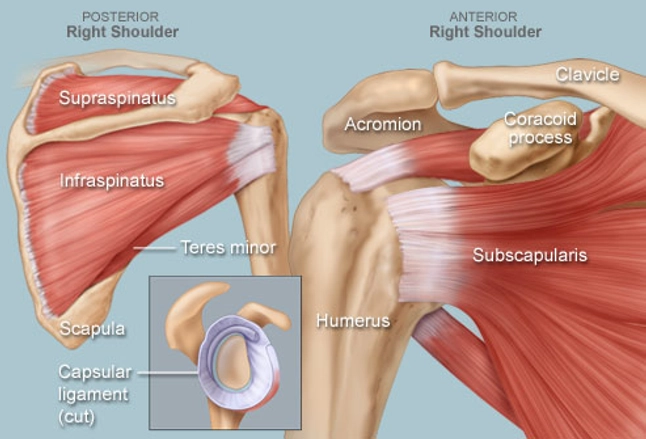
 Rotator Cuff Anatomy Posterior Download Scientific Diagram
 Shoulder Arm Atlas Of Anatomy
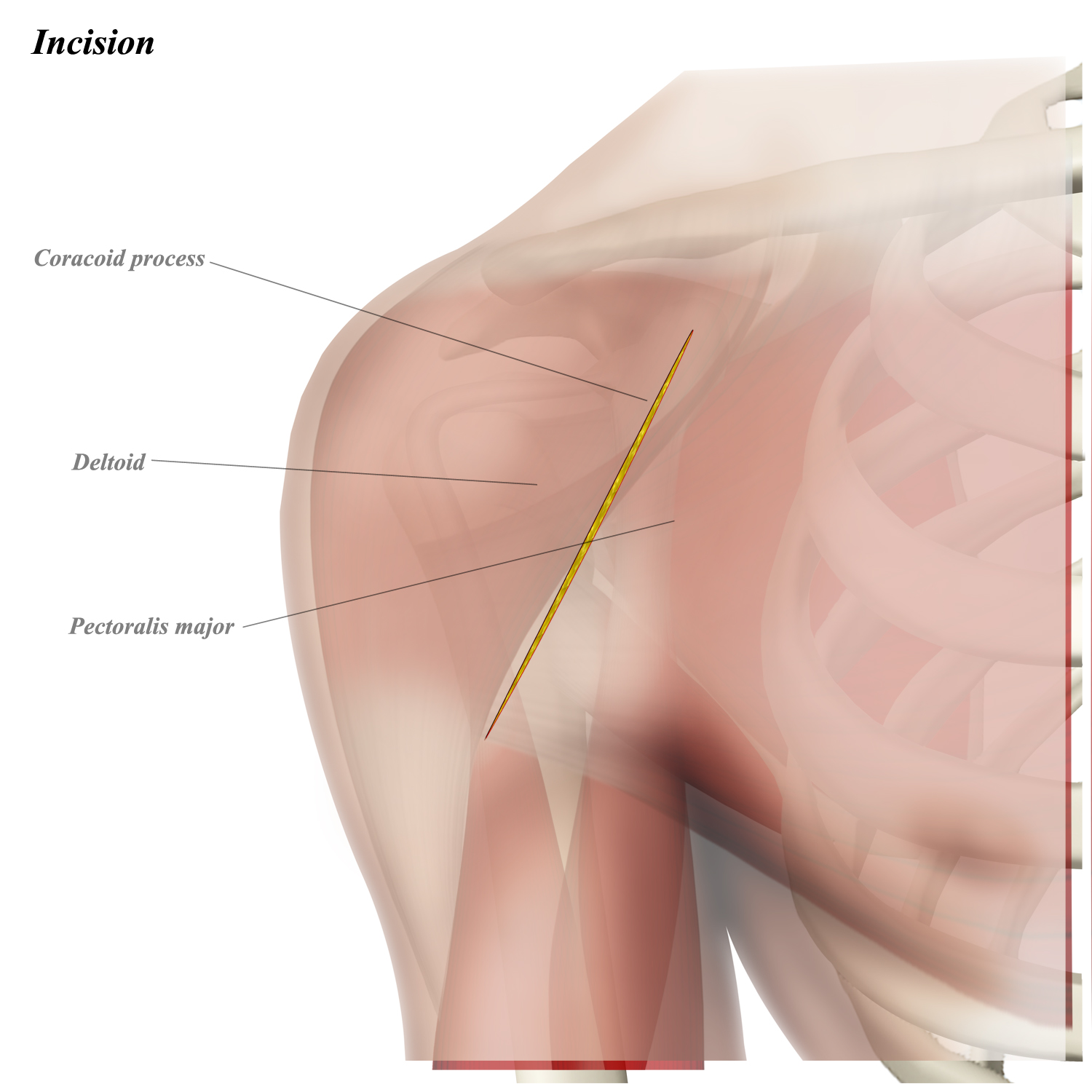 Shoulder Anterior Deltopectoral Approach Approaches
 Crossfit Shoulder Muscles Part 2 Posterior Musculature
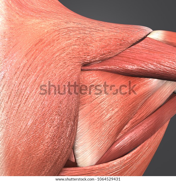 Shoulder Muscles Anatomy Posterior View 3d Stock
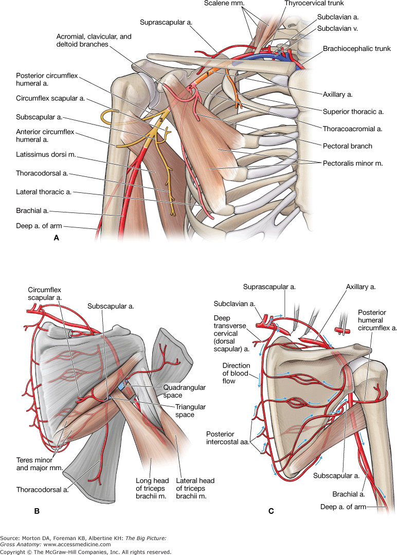 Chapter 30 Shoulder And Axilla The Big Picture Gross
 Rotator Cuff Muscles Anterior And Posterior Shoulder
 Muscles Advanced Anatomy 2nd Ed
 3 Posterior Shoulder Back 1
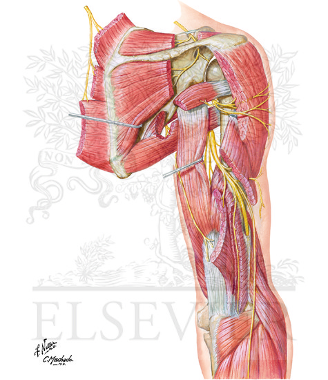 Radial Nerve In Arm And Nerves Of Posterior Shoulder

 Posterior Shoulder Joint Muscles Purposegames

 Shoulder Human Anatomy Image Function Parts And More
Belum ada Komentar untuk "Shoulder Anatomy Posterior"
Posting Komentar