Atlas And Axis Anatomy
It allows the head turn from side to side. Together they support the skull facilitate neck movement and protect the spinal cord.
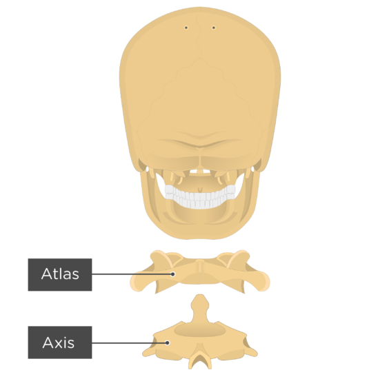
 Figure Cervical Vertebra Cervical Axis Atlas
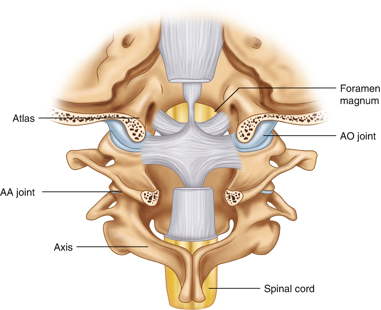
By the atlanto axial joint it forms the pivot upon which the first cervical vertebra the atlas which carries the head rotates.
Atlas and axis anatomy. This video is about many structures from the neck anatomy and gives you information about the anatomy of the atlas and axis as well as head anatomy and articular surfaces in atlas anatomy. The atlas and axis are specialized to allow a greater range of motion than normal vertebrae. Explore and learn about the second cervical vertebra called the axis with our 3d interactive anatomy atlas.
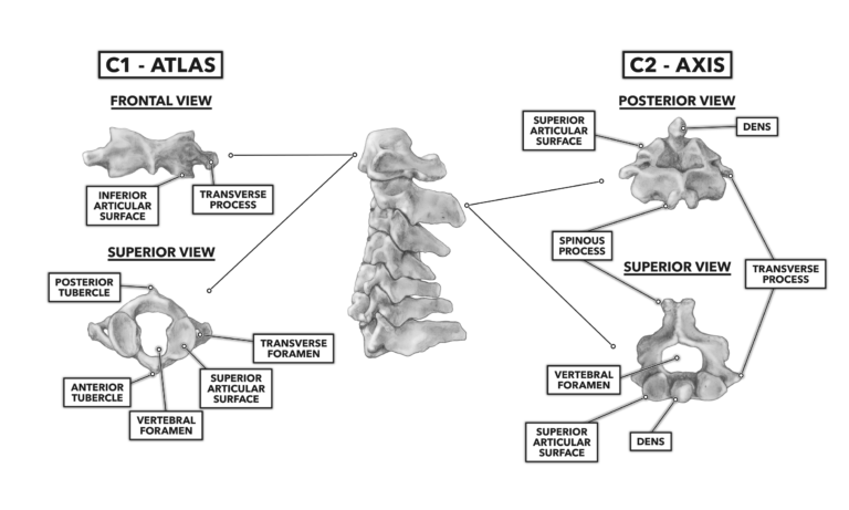
The joint between the atlas and axis is a pivot type of joint. The atlas is the topmost vertebra and with the axis forms the joint connecting the skull and spine. It is best to understand these parts by learning that the atlas vertebrae is for stability while the axis is for motion and function of the latter.
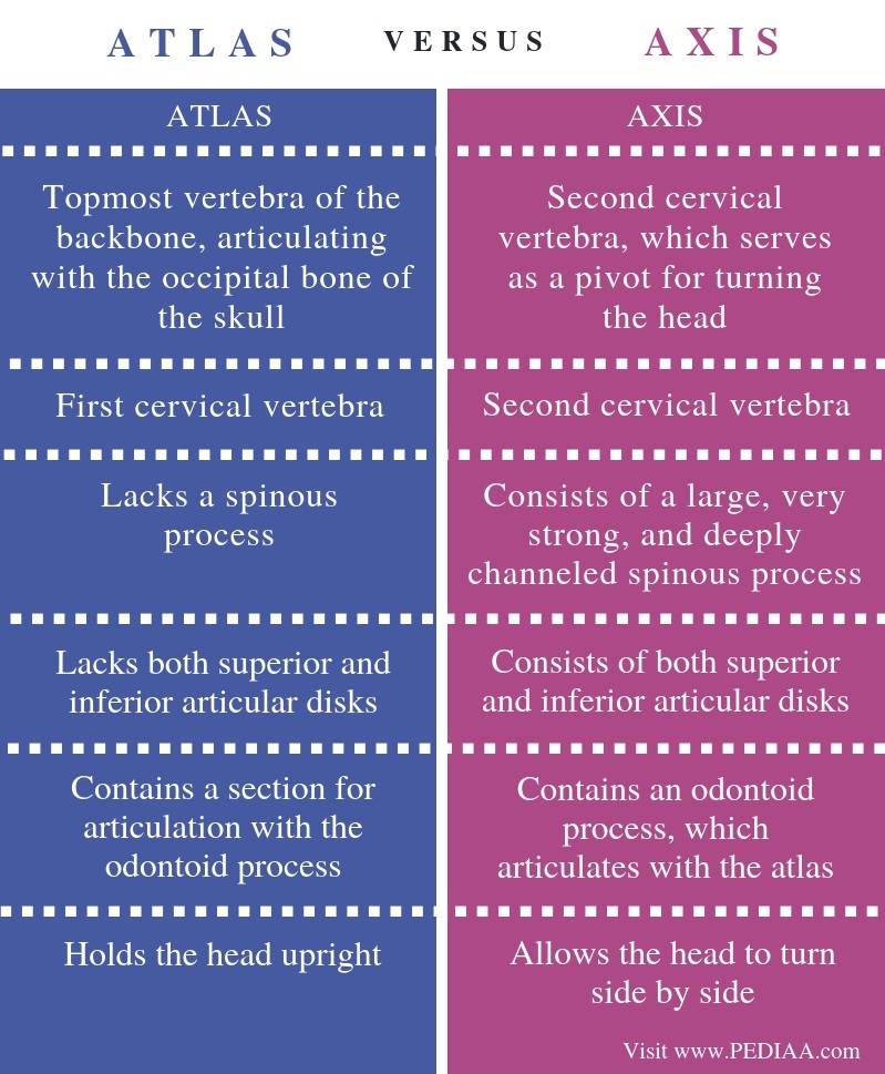
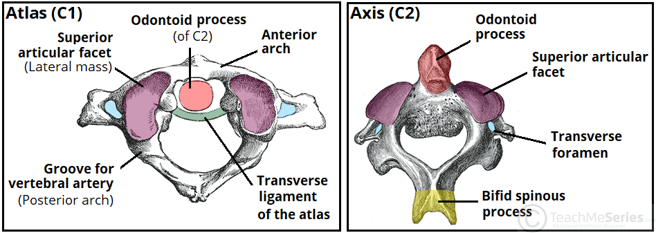
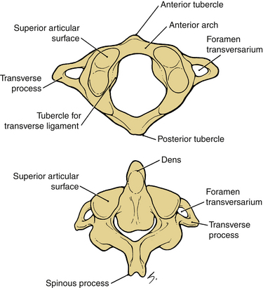
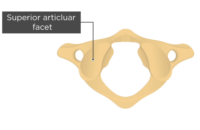
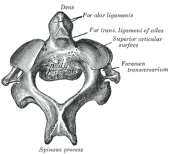
The key difference between atlas and axis vertebrae is that the atlas vertebra is the most superior vertebra. It is followed by the axis. It has what is called the odontoid process about which the atlas rotates.
The atlas and axis vertebrae are important in the balance of the skeletal frame of the human body. The atlas and axis vertebrae are the two most superior bones in the vertebral column and they are part of the seven cervical vertebrae. Axis vertebra is the second most superior vertebra of the vertebral column.
The axis is the second cervical vertebra. Without each part of the vertebrae the body cannot function properly. In anatomy the second cervical vertebra c2 of the spine is named the axis from latin axis axle or epistropheus.
It holds the head upright. The atlas is the top most bone sitting just below the skull. An interactive quiz covering atlas bone anatomy through multiple choice questions and featuring the iconic gbs illustrations.
They are responsible for the nodding and rotation movements of the head. Join us in this video where we show the anatomy of atlas and axis c1 c2 through the use of models. Please support us patreon https.
Atlas and axis vertebrae are two cervical vertebrae of the vertebral column.
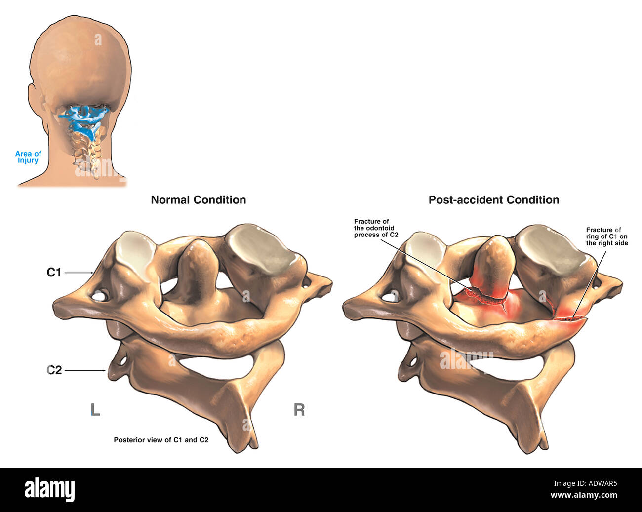
 First Cervical Vertebra An Overview Sciencedirect Topics
 The Atlas Of An Elephant Cranial Intelligence Blog
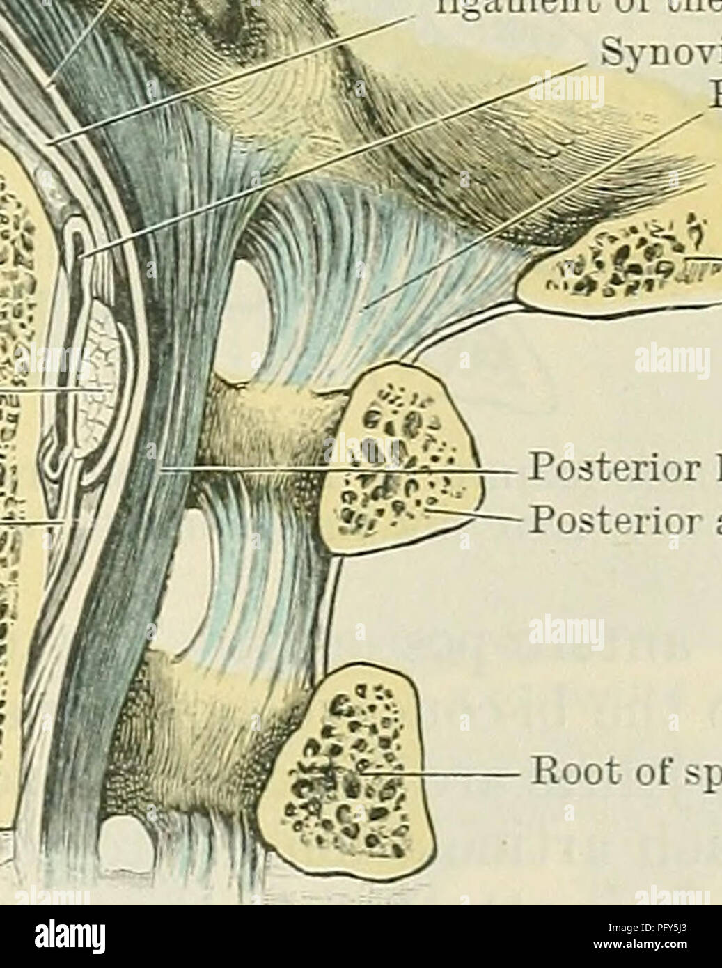 Cunningham S Text Book Of Anatomy Anatomy Aeticulation Of
 Anatomical Teaching Models Plastic Vertebrae Model Atlas
 C1 Vertebra Atlas And Accompanying Structures The Art Of
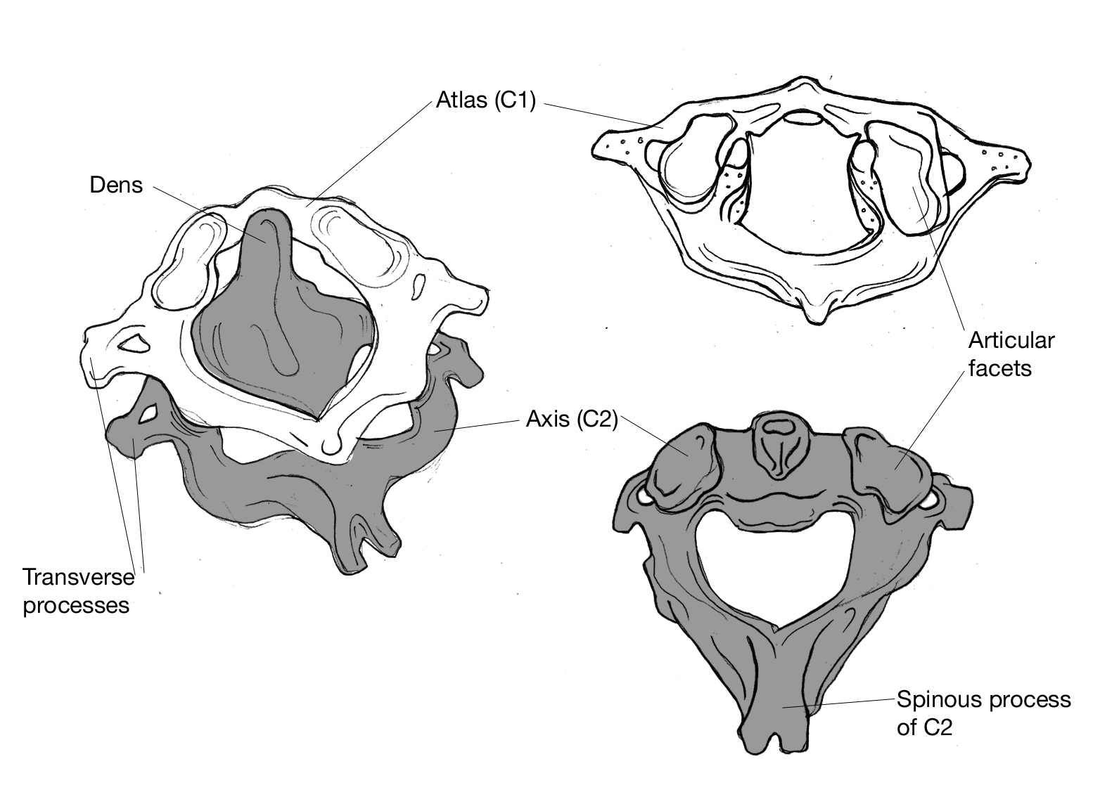
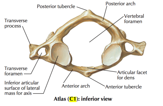
 Labelled Images Of Atlas And Axis Cervical Vertebraes Side
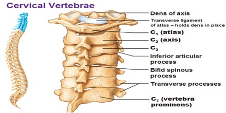
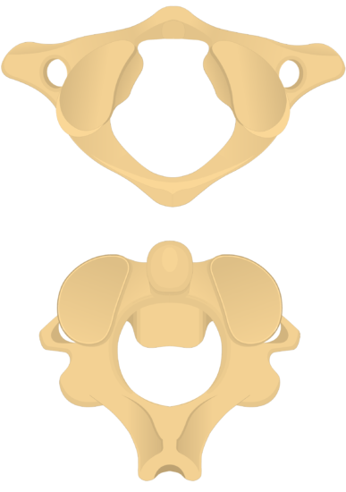
 Cervical Vertebrae Atlas Axis
 Axis Bone Anatomy
 Atlas Bone Anatomy
 Anatomy Of Vertebra Column Mbbs Batch 19 Year 1
 Atlas Vertebra Stock Photos Atlas Vertebra Stock Images
 Vector Illustration Of Cervical Vertebrae Medical Scheme With
---1st-Cervical-Vertebra.png) C1 Atlas 1st Cervical Vertebra
C1 Atlas 1st Cervical Vertebra
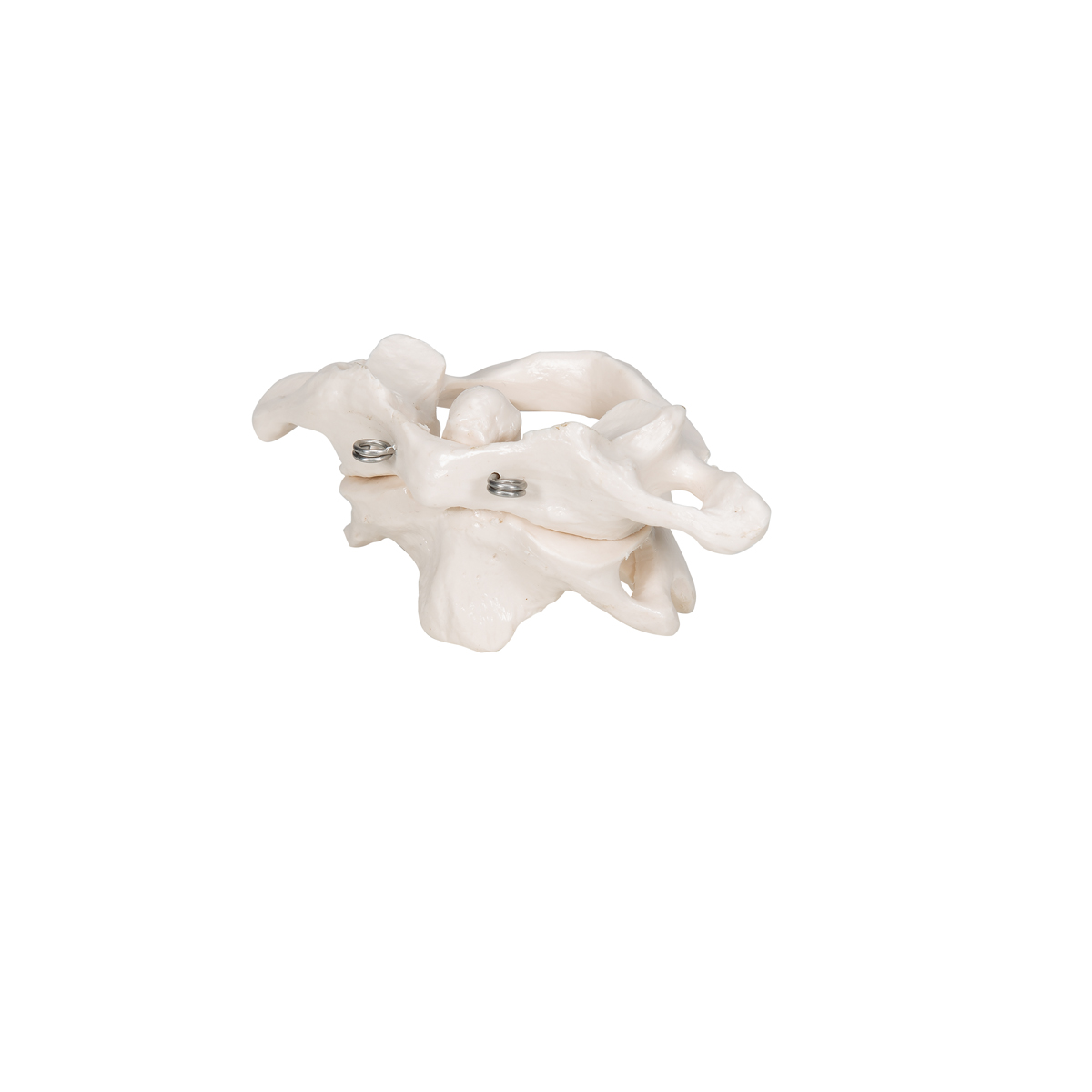
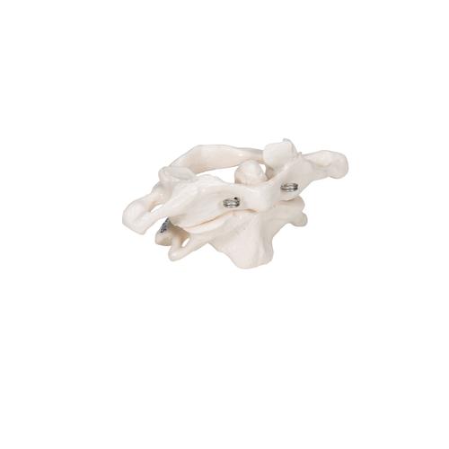 Human Atlas Axis Model Wire Mounted 3b Smart Anatomy
 Difference Between Atlas And Axis Vertebrae Compare The
 Atlantoaxial Joint Pain Springerlink
 The Cervical Spine Features Joints Ligaments
 Occipitocervical Region Clinical Gate
 Atlas Bone Anatomy
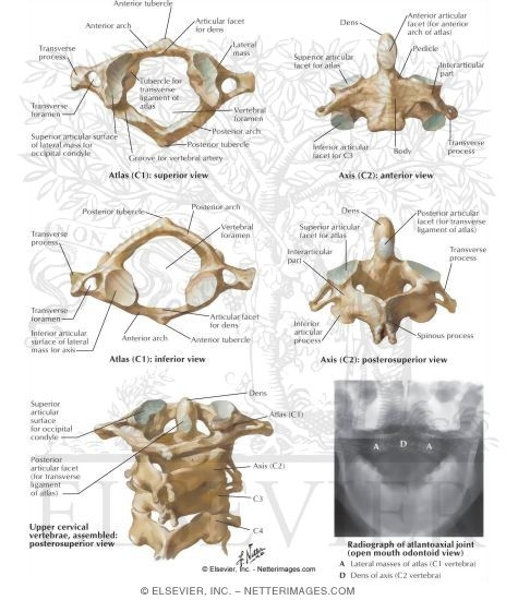
 Pediagenosis
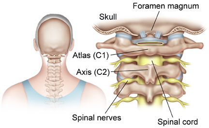
 Upper Cervical Spine Disorders Anatomy Of The Head And
 Axis Anatomy Wikipedia
 General Anatomy Of The Bull And The Cow Illustrated Atlas
 Crossfit The Cervical Vertebrae
Cervical Vertebrae Atlas And Axis Spine Osteology
Belum ada Komentar untuk "Atlas And Axis Anatomy"
Posting Komentar