Eye Diagram Anatomy
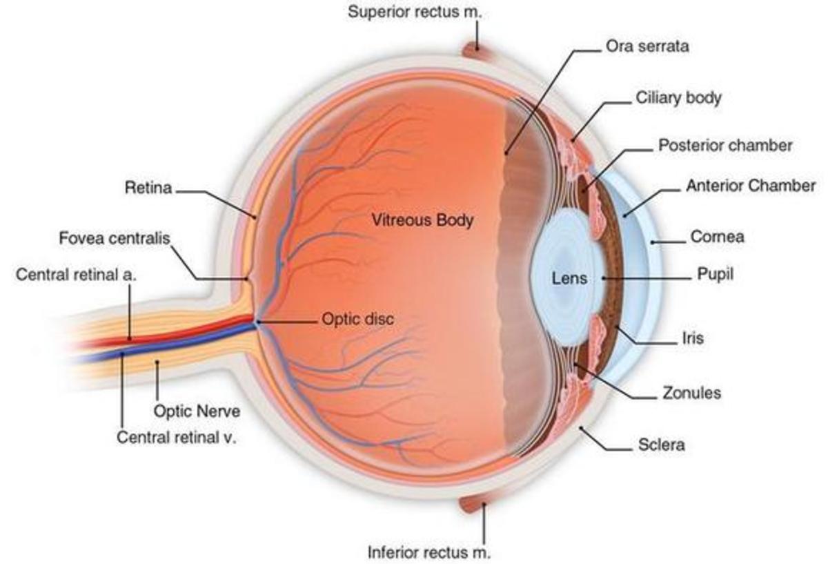
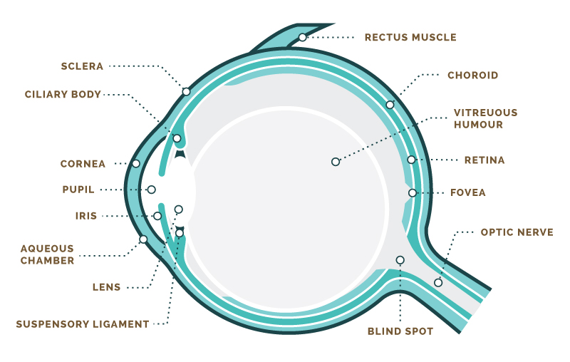
It is located opposite to the anterior chamber at the back of the lens. The eye is surrounded by the orbital bones and is cushioned by pads of fat within the orbital socket.
 Human Eye Anatomy Classroom Diagram Educational Chart Cool Wall Decor Art Print Poster 24x36
A closer look at the parts of the eye by liz segre when surveyed about the five senses sight hearing taste smell and touch people consistently report that their eyesight is the mode of perception they value and fear losing most.
Eye diagram anatomy. In biology human biology physics and practical courses in medicine nursing and therapies. The anatomy and physiology of the human eye is an important part of many courses eg. Here is a tour of the eye starting from the outside going in through the front and working to the back.
The human eye is an organ that reacts to light and has several purposesthe eye is a complex structure with layers lens muscles receptors that is surrounded by many boneswatch various parts. To understand the diseases and conditions that can affect the eye it helps to understand basic eye anatomy. Next light passes through the lens a clear inner part of the eye.
Six extraocular muscles in the orbit are attached to the eye. The iris the colored part of the eye controls how much light the pupil lets in. The eye sits in a protective bony socket called the orbit.
Learn vocabulary terms and more with flashcards games and other study tools. Learn about their function and problems that can affect the eyes. This simple introduction the subjects of the eye and visual optics includes a simple diagram of the eye together with definitions of the parts of the eye.
It is filled with a fluid called vitreous humour. By smac17 plays quiz updated nov 13 2017. Cover the alphabet us state capitals 509.
Science quiz human eye anatomy random science or biology quiz can you locate the parts of the human eye. The posterior chamber is also referred to as the vitreous body as indicated in the diagram below anatomy of the eye. Start studying eye anatomy.
The cornea is shaped like a dome and bends light to help the eye focus. Webmds eyes anatomy pages provide a detailed picture and definition of the human eyes. Extraocular muscles help move the eye in different directions.
Anatomy of the eye. The posterior chamber is a larger area than the anterior chamber. Some of this light enters the eye through an opening called the pupil pyoo pul.
Nerve signals that contain visual information are transmitted through the optic nerve to the brain.
:background_color(FFFFFF):format(jpeg)/images/library/7787/overview-of-eyeball_english.jpg) Blood Vessels And Nerves Of The Eye Anatomy Kenhub
Blood Vessels And Nerves Of The Eye Anatomy Kenhub
 Pin By Sallie Forrester On Home School General Eye
 Anatomy Of The Eye Human Eye Anatomy Owlcation
Eye Anatomy Diagram Enchantedlearning Com
Eye Anatomy And Vision Course Hero
 Human Eye Anatomy Structure And Function
 Anatomy Of The Eye Biology For Majors Ii
Anatomy Of The Eye The Johns Hopkins Wilmer Eye Institute
 Eye Diagram Photos Royalty Free Images Graphics Vectors
 Anatomy Of The Eye Red Rover Ventures
/GettyImages-695204442-b9320f82932c49bcac765167b95f4af6.jpg) Structure And Function Of The Human Eye
Structure And Function Of The Human Eye
 Human Eye Anatomy Parts Of The Eye Explained Eye Anatomy
Label Eye Printout Enchantedlearning Com
 Eye Diagram And Eye Anatomy How Vision Works

Large Human Eye Anatomy Diagram The Eye Si Gh T
 Eye Diagram Without Labels Via Anatomy Pictures Gallery If
 Amazon Com Human Eye Anatomy Medical Chart Educational
Eye Health Anatomy Of The Eye Visionaware
 Nvcc Bio 141 Ch 16 Anatomy Of Internal Eye Diagram Quizlet
 Anatomy Of The Eye Moorfields Eye Hospital
Gross Anatomy Of The Eye By Helga Kolb Webvision
 Human Anatomy Eye Diagram Macular Degeneration Pinterest
 What Is The Purpose Of Eye Diagrams Quora
 Eye Diagram Free Eye Diagram Template
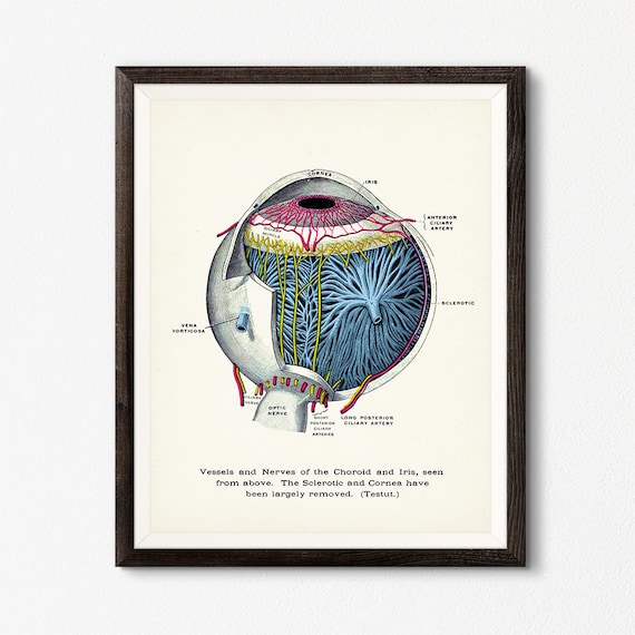 Human Eye Diagram Digital Download Optometrist Gift Eye Anatomy Poster Optometry Decor Clinic Wall Art Eye Diagram Eye Ball Art


Belum ada Komentar untuk "Eye Diagram Anatomy"
Posting Komentar