Hip X Ray Anatomy
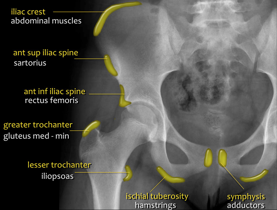
The acetabulum is formed by the three bones of the pelvis the ischium ilium and pubis. The hip joint is a ball and socket joint that represents the articulation of the bones of the lower limb and the axial skeleton spine and pelvis.
 The Radiology Assistant Hip Pathology In Children
The distance between the x ray tube and the film should be 12 m.

Hip x ray anatomy. The horizontal beam lateral hip radiograph or shoot through hip is the in the purist terms the orthogonal view of the neck of the femur to the ap projection 13. Only an x ray was taken no mri. Please click on the thumbnail image to launch a full sized image that is annotated with the correct landmarks.
Pelvic and hip x rays are most frequently obtained when there is concern for fracture joint dislocation and effusion and several pediatric pathologies involving the pelvic girdle which are outlined below. Years went by as i complained of butt pain when i sat anywhere. A standard hip x ray examination generally includes an anteroposterior pa image and a lateral image.
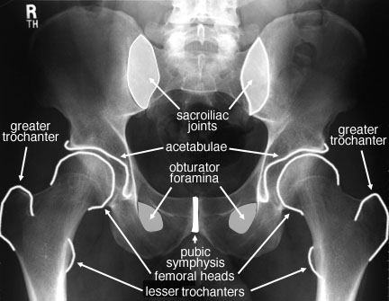
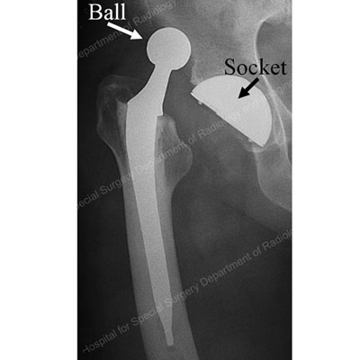
Front view of the hip joint bones. Xray anatomy of the hip xray anatomy of the hip here are a series of xrays that are illustrated and annotated to identify the anatomic landmarks and concepts that are used during total hip arthroplasty. Hip x ray anatomy normal ap shentons line is formed by the medial edge of the femoral neck and the inferior edge of the superior pubic ramus loss of contour of shentons line is a sign of a fractured neck of femur important note.
An anteroposterior hip radiograph includes images of both sides of the hip on the same film and projects towards the middle of the line connecting the upper symphysis pubis and anterior superior iliac spine. The projection is used to assess the neck of the femur in profile during the investigation of a suspected neck of femur fracture 2. Hip anatomy function and common problems.
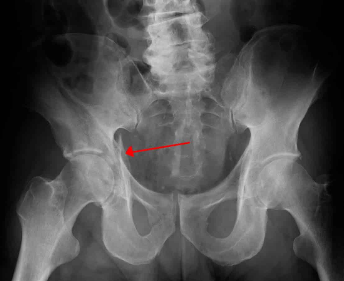
Anatomy of the hip. I was told i had a l pelvic avulsion fracture n was sent home on crutches n told itll heal. The rounded femoral head sits within the cup shaped acetabulum.
Fractures of the femoral neck do not always cause loss of shentons line. The lateral direction may be opted for in axiolateral images or a frog leg lateral image. Before delving into the radiographic approach to pelvic and hip x rays let us first review some anatomy.
Ideally the ap image shows both hip joints which strictly speaking makes it a pelvis x ray to allow comparison with the other hip. In plain radiography x ray anteroposterior and lateral hip radiographs are usually taken.
 How To Read Pelvic X Rays International Emergency Medicine
 Startradiology

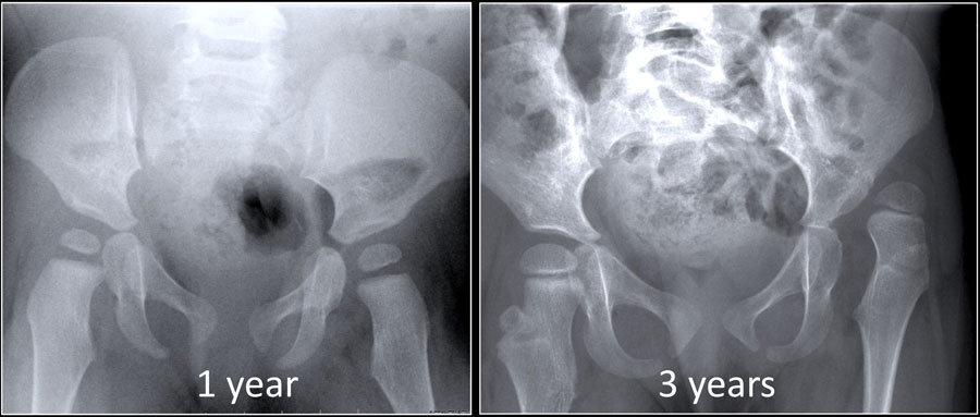
 Pelvic X Ray Normal Different Ages Radiology Case
 Film Critique Of The Lower Extremity Part 1
 Femoral Neck Fractures Trauma Orthobullets
 International Hip Dysplasia Institute
X Ray Film Reading Made Easy
 Back To Basics Pelvic Xrays Taming The Sru
 Film Critique Of The Lower Extremity Part 1
 Normal Radiographic Anatomy Of The Hip Radiology Case
 Startradiology
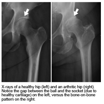 Treating Hip Arthritis Mu Health Care
 The Hip Bone Ilium Ischium Pubis Teachmeanatomy
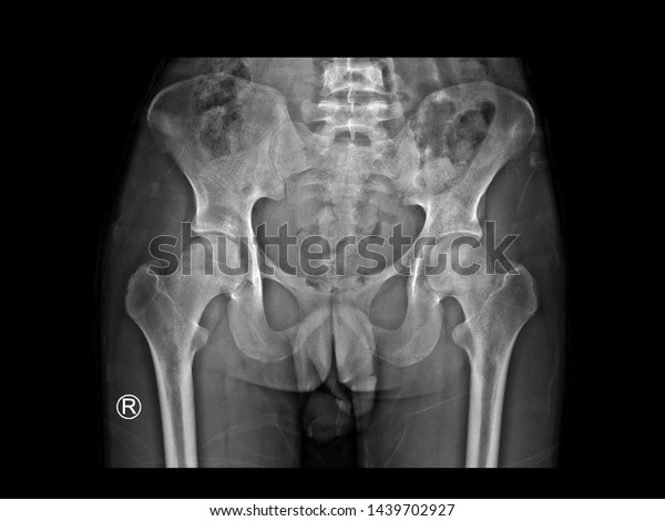 Film X Ray Hip Radiograph Show Stock Photo Edit Now 1439702927
 Radiological Anatomy Of The Lower Limb

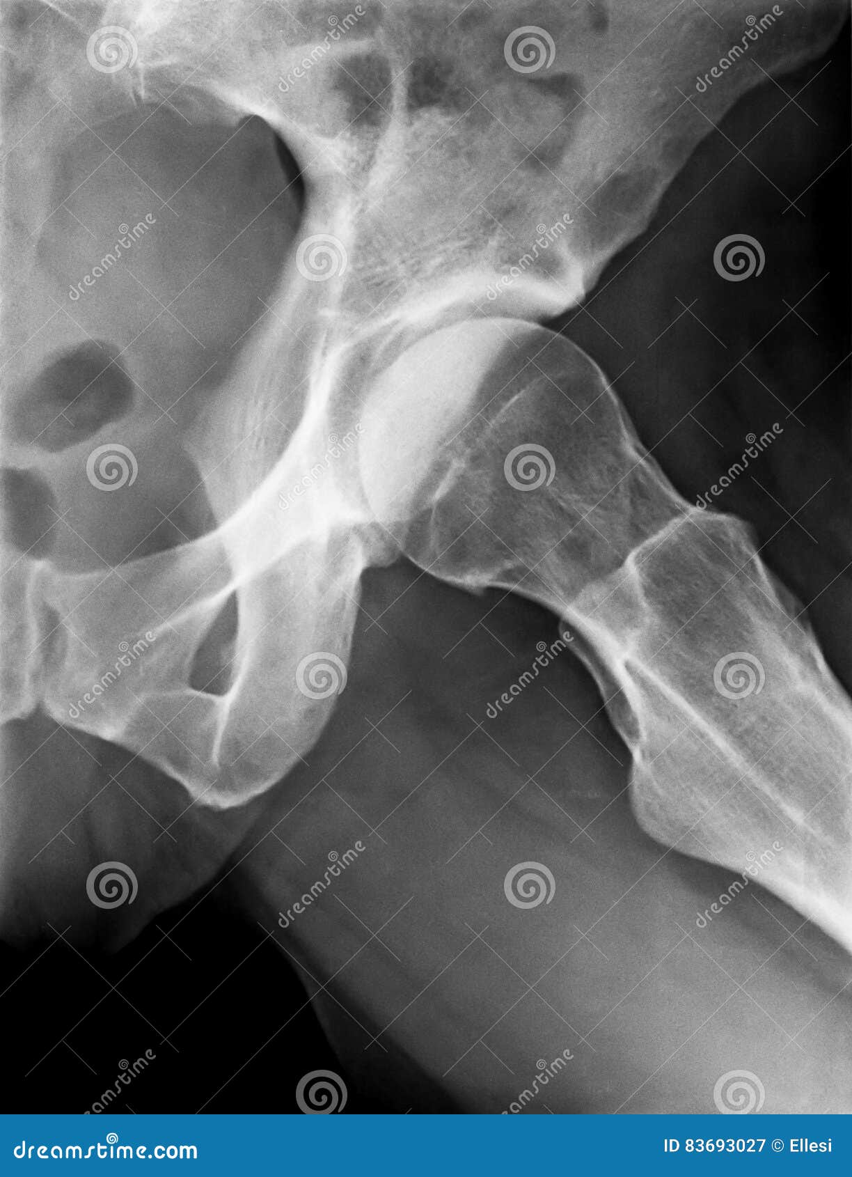 X Ray Of Female Left Hip Stock Image Image Of Anatomy
Knee X Rays
 The Radiology Assistant Developmental Dysplasia Of The Hip
 Knee X Rays
 Dislocated Hip Symptoms Diagnosis And Treatments Hss
 Untitled Document
 Ilium Bone Hip Bone Image Photo Free Trial Bigstock
Belum ada Komentar untuk "Hip X Ray Anatomy"
Posting Komentar