Anatomy Of The Knee Nerves
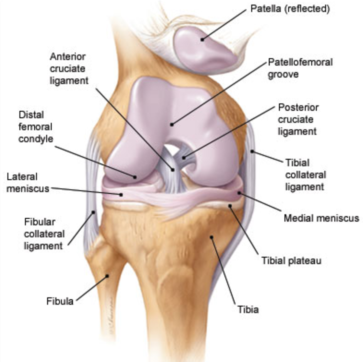
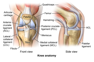
Helps to lower and raise the body. The anterior cruciate ligament prevents the femur from sliding backward on the tibia or the tibia sliding forward on the femur.
 Anatomy Of The Knee Bones Muscles Arteries Veins Nerves
The anatomy of the knee knee bones knee muscles knee arteries knee veins and nerves looking into the anatomy of the knee.

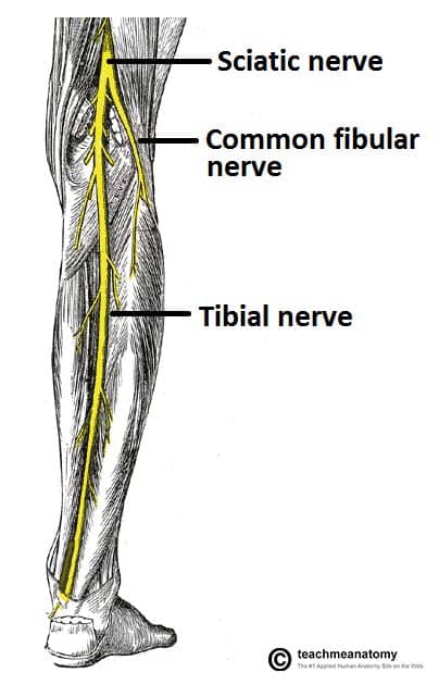
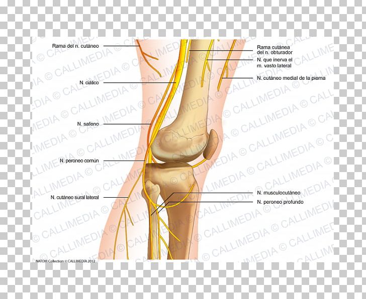
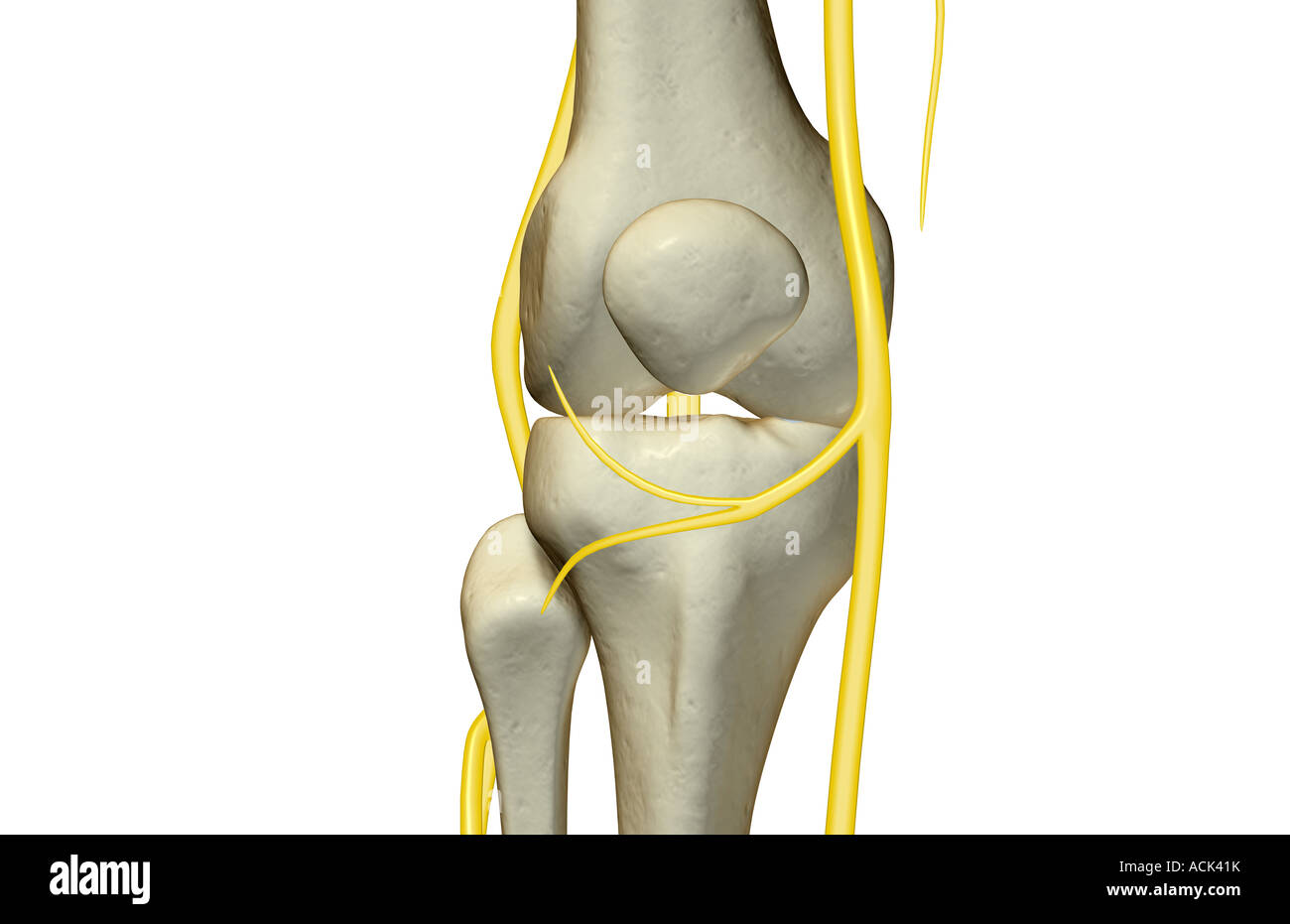
Anatomy of the knee nerves. The common peroneal nerve diverges laterally running just behind the tendon of biceps femoris. The most important nerves around the knee are the tibial nerve and the common peroneal nerve in the back of the knee. The sciatic nerve travels down the thigh to the area of the popliteal fossa and at this point it divides into the tibial and common peroneal nerves.
The knee is designed to fulfill a number of functions. Above the knee the sciatic nerve divides into two major nerves the tibial nerve and the common peroneal nerve. The large sciatic nerve splits just above the knee to form the tibial nerve and the common peroneal nerve.
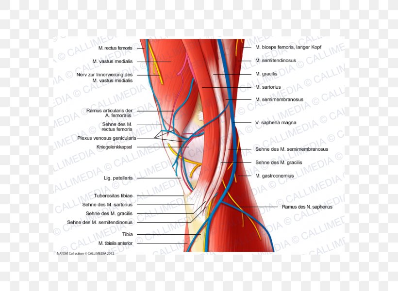
Medial sural cutaneous nerve. Infrapatellar br of saphenous nerve medial crural cutaneous nerve cutaneous br of obturator nerve saphenous nerve articular br of obturator nerve to knee posterior femoral cutaneous nerve tibial nerve medial sural cutaneous nerve common fibular nerve sural nerve lateral sural cutaneous nerve deep fibular nerve superficial. Common fibular peroneal nerve.
These two nerves travel to the lower leg and foot supplying sensation and muscle control. Helps propel the body forward. Ligaments join the knee bones and provide stability to the knee.
Acts as a shock absorber. The most important nerves around the knee are the tibial nerve and the common peroneal nerve in the back of the knee. The popliteal fossa is a closely packed space.
Support the body in an upright position without the need for muscles to work. These two nerves travel to the lower leg and foot supplying sensation and muscle control. The tibial nerve runs downward in the midline and passes between the two heads of gastrocnemius along with the popliteal vessels.
Makes walking more efficient. Full list of the names of bones muscles veins arteries and veins found in the knee. This nerve branches off the tibial nerve.
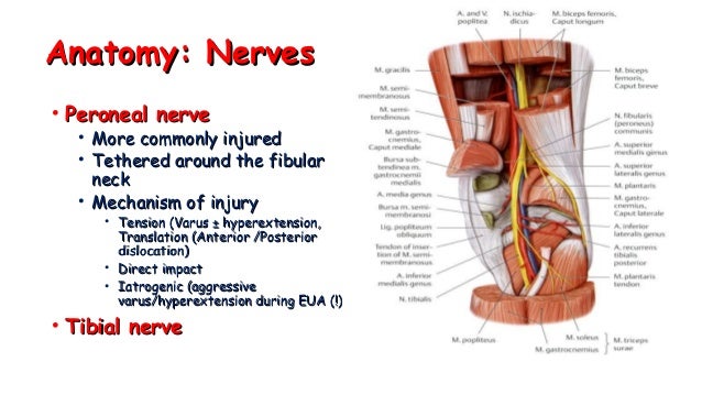
This nerve branches off the sciatic nerve in the popliteal fossa and runs along the biceps femoris and leaves the fossa to run around the head of the fibula and down the leg to the ankle. Allows twisting of the leg.
 Distal Femur Reduction Fixation Orif Dynamic
 The Common Fibular Nerve Course Motor Sensory
Knee Injury Types Symptoms Exercises Treatment Diagnosis
 Core Anatomy Winding Round To Foot Drop Which Nerve Is
 Uncommon Injuries Posterior Interosseous Nerve Dysfunction
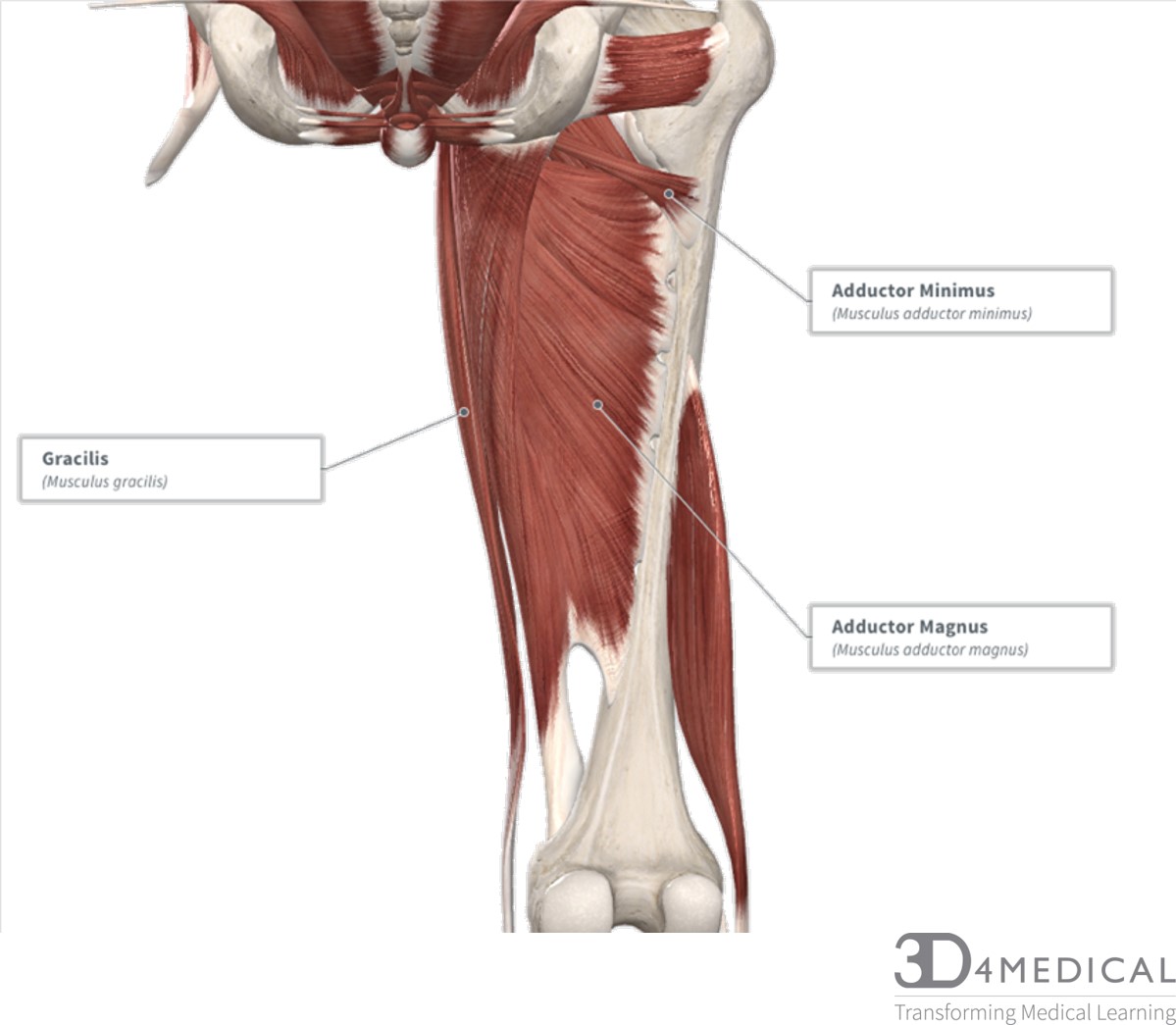 Muscles Advanced Anatomy 2nd Ed

 Anatomy Of The Saphenous Nerve Download Scientific Diagram
 01 Knee Anatomy
 Anatomy Of The Saphenous Nerve Doctor Stock
 Thumb Common Peroneal Nerve Knee Human Anatomy Png Clipart
 Human Leg Nerves Stock Photos Human Leg Nerves Stock
 Ultrasound Guided Obturator Nerve Block Nysora
 Surface Anatomy Advanced Anatomy 2nd Ed
 Neurovasculature Of The Leg And Knee Region Preview Human Anatomy Kenhub

 Knee Wikipedia
 Sciatic Nerve An Overview Sciencedirect Topics
 Nerves Knee Stock Photos Nerves Knee Stock Images Alamy
 Nerve Muscle Medial Knee Injuries Human Anatomy Png
 Saphenous Nerve Block Overview Indications Contraindications
 Femoral Nerve Physiopedia
 Patella Approach Mid Axial Longitudinal Approach Ao
![]() Leg And Knee Anatomy Bones Muscles Soft Tissues Kenhub
Leg And Knee Anatomy Bones Muscles Soft Tissues Kenhub
 Knee Anatomy Exhibits
 The Anatomy And Pattern Of Pain Of The Saphenous Nerve At
Belum ada Komentar untuk "Anatomy Of The Knee Nerves"
Posting Komentar