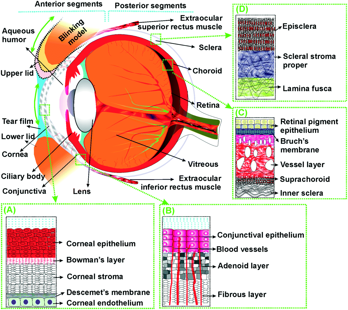
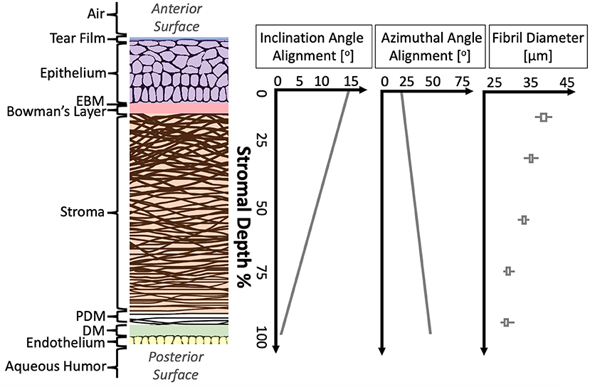
Corneal Anatomy
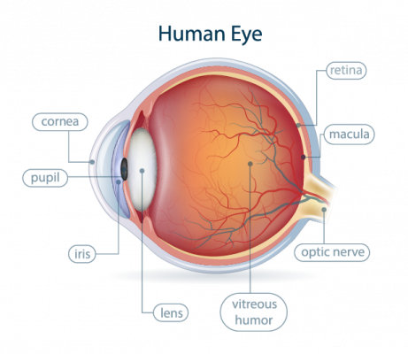
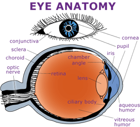
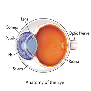
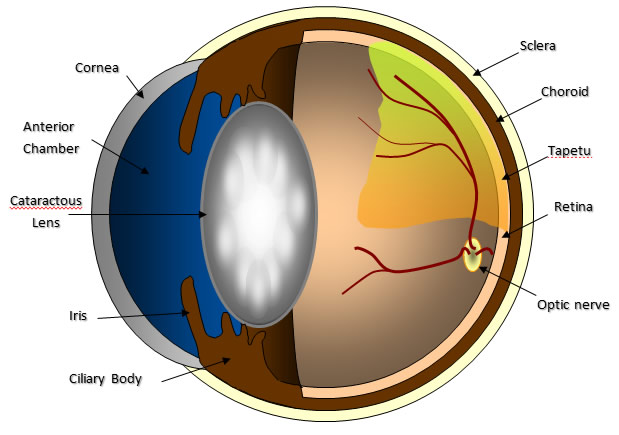
The iris of the eye functions like the diaphragm of a camera controlling the amount. It covers the pupil the opening at the center of the eye iris the colored part of the eye and anterior chamber the fluid filled inside of the eye.
 Corneal Anatomy Lightning Strikes Twice
The eyes crystalline lens is located directly behind the.
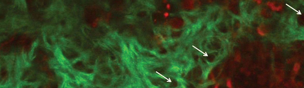
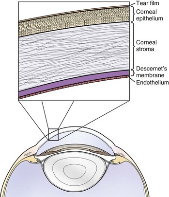
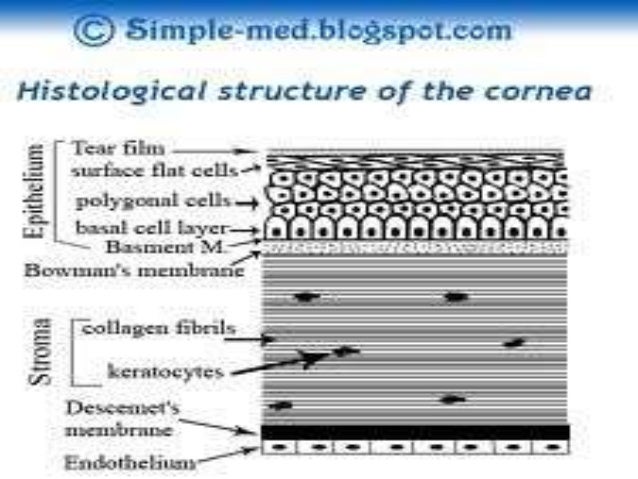
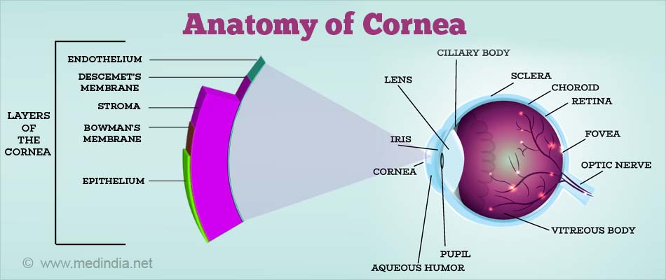
Corneal anatomy. Except at its margins the cornea contains no blood vessels but it does contain many nerves and is very sensitive to pain or touch. Transparent refract light contain the intraocular pressure provide a protective interface sbj. The cornea is the transparent part of the eye that covers the front portion of the eye.
Cornea dome shaped transparent membrane about 12 mm 05 inch in diameter that covers the front part of the eye. The cornea is a transparent structure that together with the lens provides the refractive power of the eye. Corneal anatomy the cornea is the transparent front part of the eye that covers the iris pupil and anterior chamber.
The anatomy and structure of the adult human cornea. In a number of ways the human eye works much like a digital camera. In humans the refractive power of the cornea is approximately 43 dioptres.
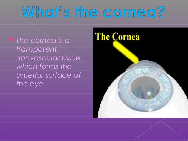
This magnified image of a section of the eye demonstrates the structure of the cornea and the limbus. The cornea the cornea is a transparent avascular tissue with a smooth convex outer surface and concave inner surface which resembles a small watch glass. Light is focused primarily by the cornea the clear front surface of the eye.
Together with the lens the cornea refracts light accounting for approximately two thirds of the eyes total optical power. The importance of the cornea to the ocular structure and visual system is often overlooked because of the corneas unassuming transparent nature. The cornea with the anterior chamber and lens refracts light with the cornea accounting for approximately two thirds of the eyes total optical power.
It is nourished and provided with oxygen anteriorly. The corneas main function is to refract or bend light. The cornea composes the outermost layer of the eye.
The cornea lacks the neurobiological sophistication of the retina and the dynamic movement of the lens. To meet the diverse functional demands the cornea must be. The cornea is the transparent front part of the eye that covers the iris pupil and anterior chamber.
Yet without its clarity the eye would not be able to perform its necessary functions.

 Eye Anatomy Detail Picture Image On Medicinenet Com
 Anatomy Of The Cornea A Section Of The Anterior Part Of
 The Anatomy Of The Frog Frogs Anatomy Amphibians
 Evaluation And Management Of Corneal Abrasions American
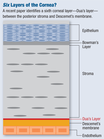
More Details On Dua S Layer Of The Cornea
 Microengineered Biomimetic Ocular Models For
 Anatomy Spokane Eye Clinic
Corneal Anatomy Symptoms And Examination American
 Eye Anatomy Glaucoma Research Foundation
 Frontiers A Review Of Structural And Biomechanical Changes

 Corneal Ulcers Veterian Key

 Cornea Definition And Detailed Illustration
 Understanding Equine Vision And Eye Disease Horse Journals
 Anatomy Of The Cornea


 Rosh Review Corneal Ulcer Optometry Eye Anatomy
 Wikipremed
 Corneal Anatomy Arya
 Anatomy Of The Eye Hummel Eye Associates Oklahoma City

 Keratitis A Clinical Approach Intechopen
 Cornea
 The Cornea Ocular Surface Center Berlin
 The Cornea Ocular Surface Center Berlin
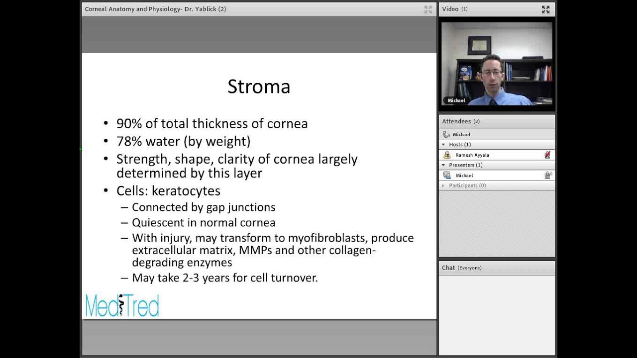 Full Lecture Corneal Anatomy And Physiology
 2a Anatomy Review Cornea Human Physiology 116 With
 Corneal Transplantation
Belum ada Komentar untuk "Corneal Anatomy"
Posting Komentar