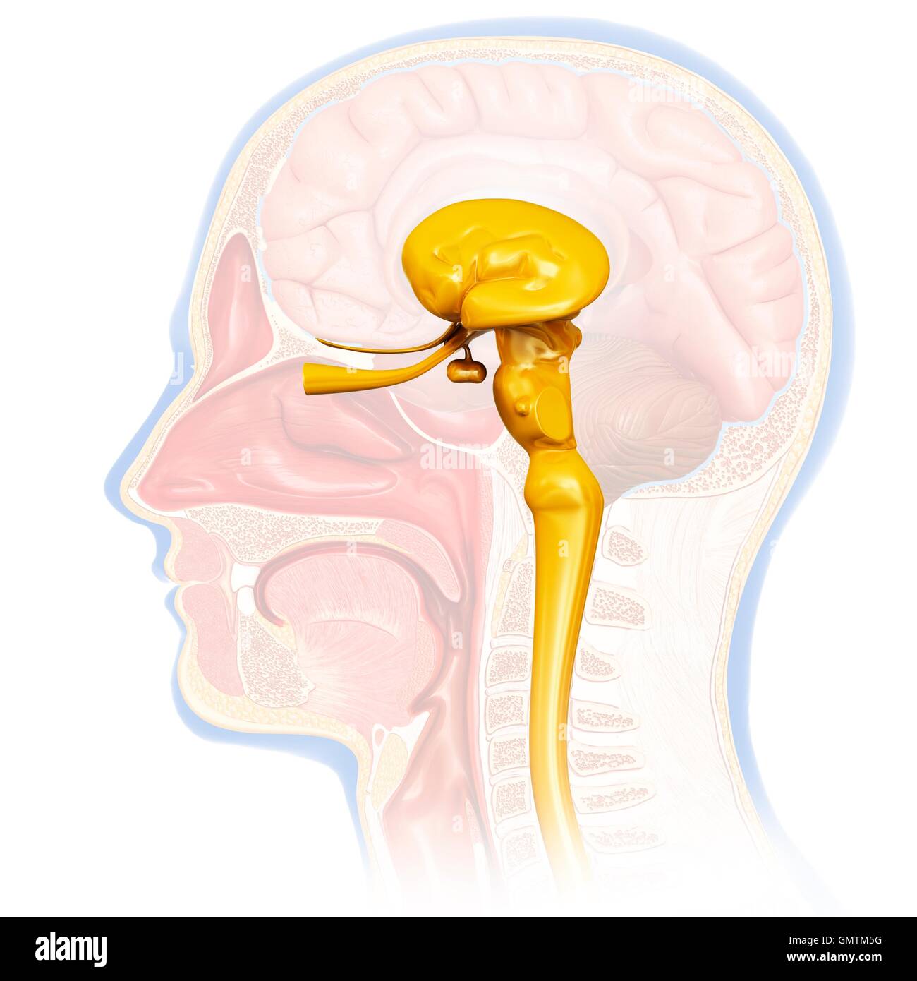
Midbrain Anatomy
There are three main parts of the midbrain the colliculi the tegmentum and the cerebral peduncles. Within the lumen of the midbrain lies the cerebral aqueduct which acts as.
 Midbrain And Pons Anatomy Location Parts Definition Kenhub
In this article we will discuss the anatomy of the midbrain its external anatomy internal anatomy and vasculature.

Midbrain anatomy. The midbrain or mesencephalon plural. The midbrain mesencephalon and the diencephalon constitute the anterior portion of the brainstem. A number of nerve tracts run through the midbrain that connect the cerebrum with the cerebellum and other hindbrain structures.
The midbrain is a portion of the brainstem positioned above the pons at the very top of the brainstem directly underneath the cerebellum. This 2 cm long structure is made up of a left and a right pedunculus cerebri or cerebral peduncles anteriorly. A major function of the midbrain is to aid in movement as well as visual and auditory processing.
The midbrain also known as the mesencephalon is the most superior of the three regions of the brainstem. The roof of the midbrain or tectum developed as the primary visual centre. The midbrain or mesencephalon is a portion of the central nervous system associated with vision hearing motor control sleepwake arousal and temperature regulation.
It acts as a conduit between the forebrain above and the pons and cerebellum below. Mesencephala or mesencephalons is the most rostral part of the brainstem and sits above the pons and is adjoined rostrally to the thalamus. The midbrain is a mesencephalic structure that extends from the superior medullary velum of the pons roof of the fourth ventricle to the posterior commissure of the third ventricle.
Midbrain structure function cranial nerve nuclei. Sensory and motor nuclei for cranial nerves extend from the hindbrain to the midbrain. The major cranial nerve nuclei within the midbrain are.
During development the midbrain forms from the middle of three vesicles that arise from the neural tube. The tectum roof has four colliculi two rostral and two caudal. Gross anatomy of the midbrain.
Midbrain is divided into two parts by an imaginary transverse line passing through the cerebral aqueduct. The mesencephalon or midbrain is the portion of the brainstem that connects the hindbrain and the forebrain. This is one of the most important components of the central nervous system cns as all neuronal transmissions that pass through the body throughout the peripheral nervous system pns to the cns are relayed must at some point to andor from the brain pass through the midbrain.
The midbrain is the topmost part of the brainstem the connection central between the brain and the spinal cord. The part ventral to the imaginary line is formed by a pair of cerebral peduncles.
 Midbrain Anatomy Parkinsons Disease Stock Illustration
 Brain Anatomy White Matter Cerebellum Cerebral Cortex
 Easy Notes On Midbrain Learn In Just 4 Minutes Earth S Lab
 Brain Stem Anatomy Location Function Anatomy Info
 The Midbrain Mesencephalon Brain Human Anatomy Course
 Midbrain Anatomy Physiology Wikivet English
 Midbrain Definition Function Structures
 Midbrain Images Stock Photos Vectors Shutterstock
 Illustration Of Human Midbrain Anatomy Stock Photo
 The Midbrain Queensland Brain Institute University Of
 Midbrain Definition Function Structures
 Stroke Medicine For Stroke Physicians And Neurologists
 Brain Anatomy Anatomy Biology Brain En Functions
 Midbrain Anatomy Images Britannica Com
 Brainstem Ii Pons And Cerebellum Part 1
 Midbrain Anatomy At The Level Of The Superior Colliculus
 Anatomical Structure Of The Midbrain Download Scientific
 Upper Midbrain Craniosacral Therapy Cranial Nerves Neurology
 Midbrain Images Stock Photos Vectors Shutterstock
 Brain Stem Midbrain Brain Stem Anatomy Study Brain
 Forebrain Midbrain Hindbrain Diagram Quizlet

Belum ada Komentar untuk "Midbrain Anatomy"
Posting Komentar