Pes Anatomy
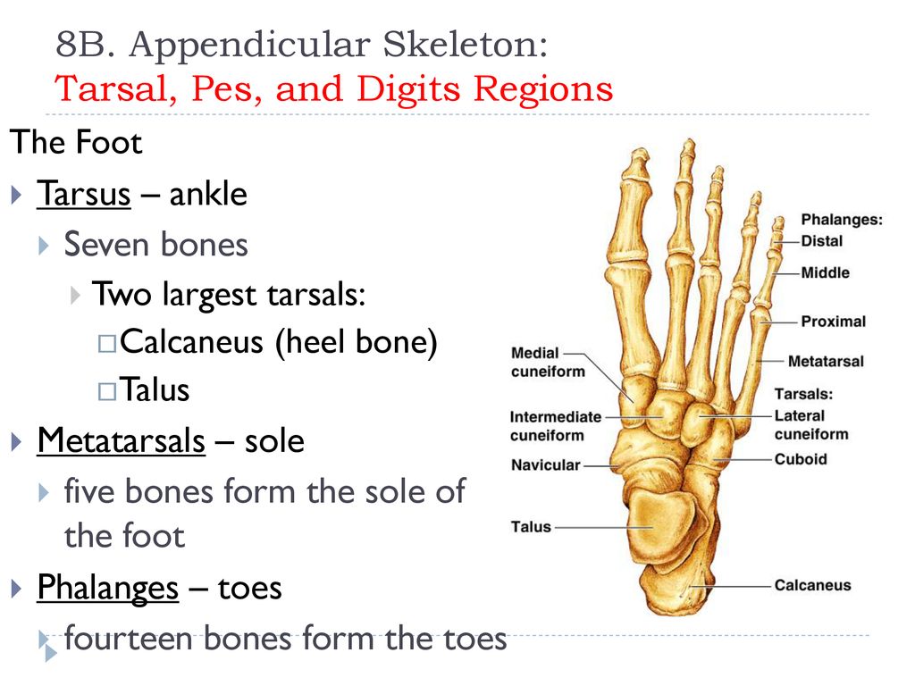
It is the part of the pentadactyl limb that includes the metatarsals and digits phalanges. To identify locate and correlate structures of the body and mark the topography by surface anatomy.
 Vector Illustration Of A Healthy Knee And Unhealthy Knee With
The tarsus metatarsus and phalanges are all encompassed within the framework of the foot mildly resembling the framework for the hand.

Pes anatomy. During evolution it has taken many forms and served a variety of functions. The part of the leg of a human being below the ankle joint. Of course they also need to be equipped to provide leverage.
Pes anatomy the pes latin for foot is the zoological term for the distal portion of the hind limb of tetrapod animals. Pes the part of the leg of a human being below the ankle joint. Despite their similarities the bones within the foot vary to indicate their ability to take the full weight of the human body.
To identify the various normal tissues under microscope differentiate and discuss them. To identify the anatomical basis of imaging mri ct scan and ultrasonography and interpret normal radiological images. Foot human foot tootsie baby talk.
His bare feet projected from his trousers. A deformity that develops after skeletal maturity is reached is commonly referred to as adult acquired flatfoot deformity aafd. Pes anserine bursitis is when there is inflammation of the pes anserine bursa causing medial knee pain.
Armored from head to foot. A bursa known as the pes anserinus bursa lies between the pes anserinus tendons and. Human human being homo man any living or extinct member of the family hominidae characterized by superior intelligence articulate speech and erect carriage.
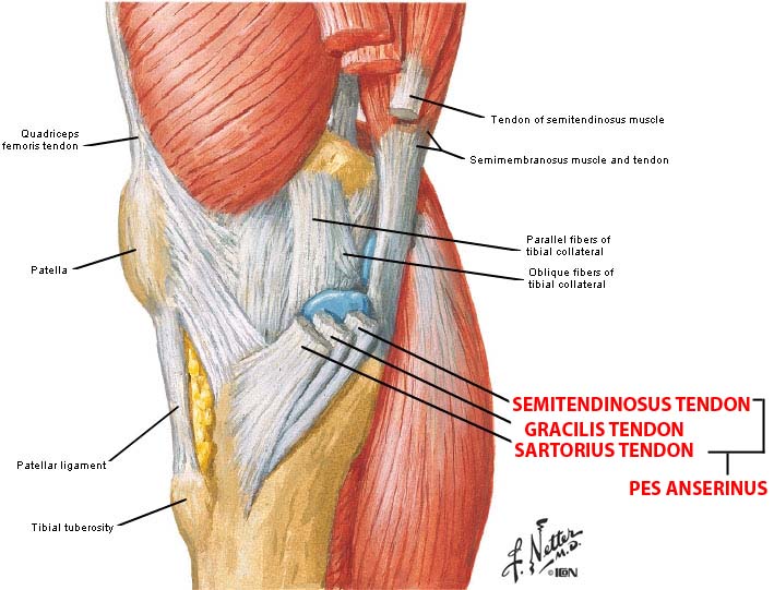
Progressive pes planus flatfoot deformity in adults is a common entity that is encountered by orthopedic surgeons. Pes general anatomy definition. The pes anserinus is an area on the medial inner side of the knee where three muscle tendons attach to the tibia shin bone.
Pes anserinus gross anatomy. We can locate this at the proximal medial aspect of the knee two inches below the medial knee joint line between the pes anserinus tendons912939139391493. The term pes anserinus may also be used to describe the branching.
The name comes from the latin for gooses foot in view of the similarity. Pes anserine bursitis also known as intertendinous bursa is an inflammatory condition of bursa of the conjoined insertion of the sartorius gracilis and semitendinosus1.
![]() Arches Of The Foot Anatomy Kenhub
Arches Of The Foot Anatomy Kenhub
Leg Anatomy Britannica
 Human Anatomy And Physiology Honors Unit 5 Chapter 5 Ppt
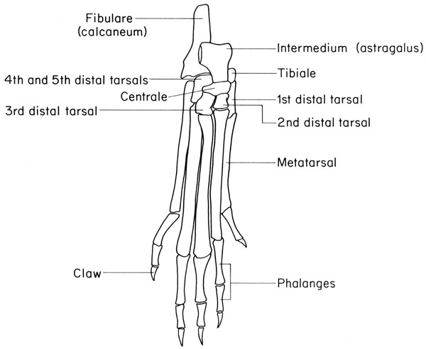
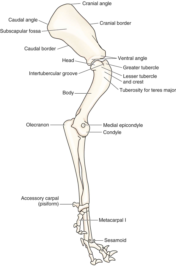
 Anatomy Of The Leg And Pes Of The Domestic Animals
 Anatomical Teaching Models Plastic Human Joint Models
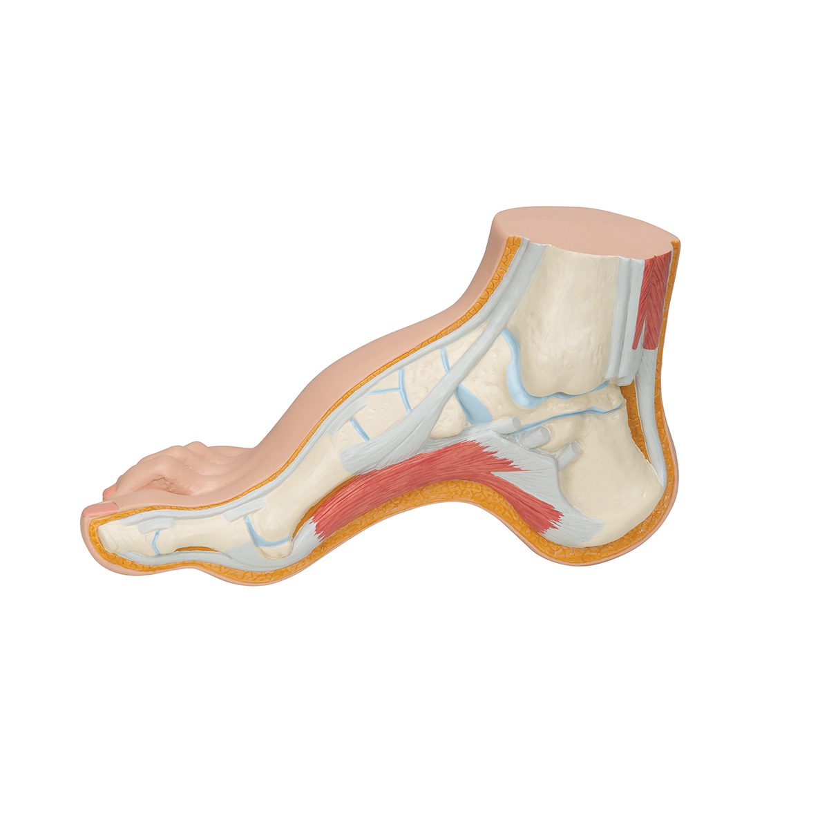
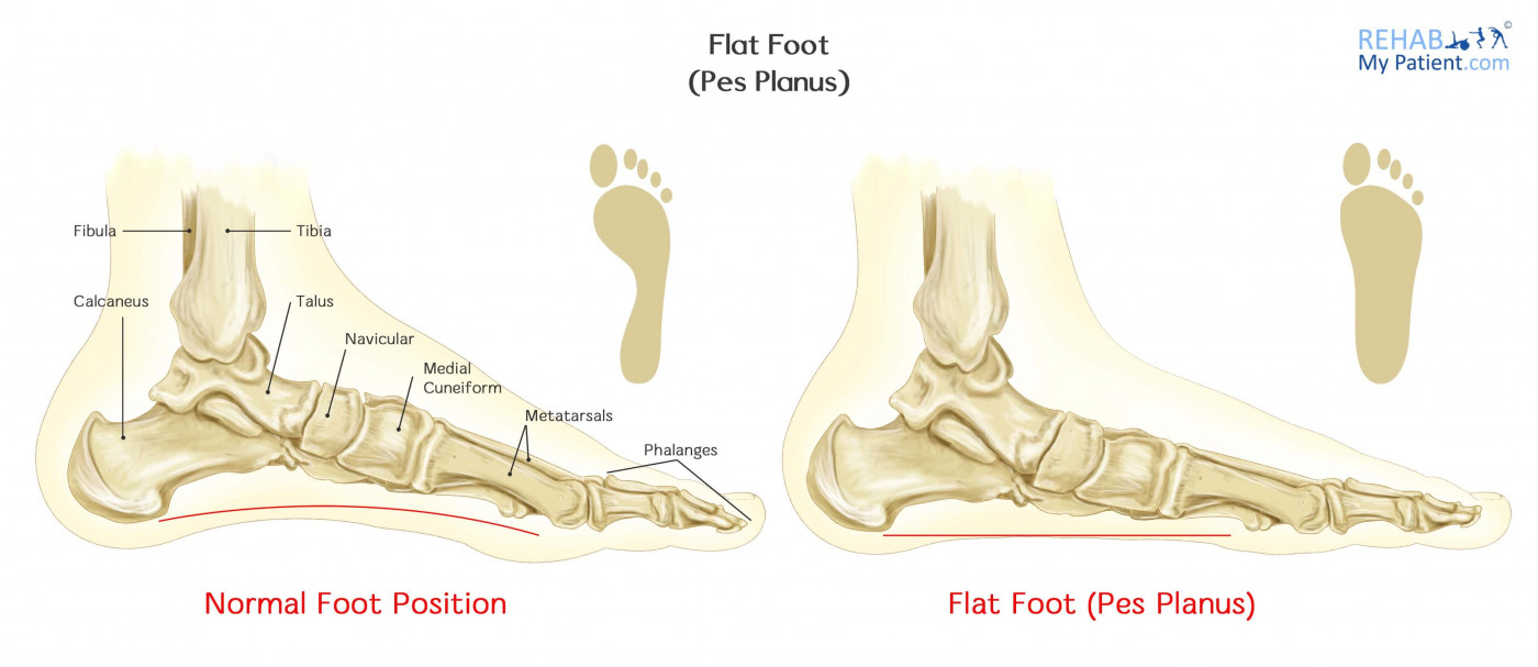 Flat Foot Pes Planus Rehab My Patient
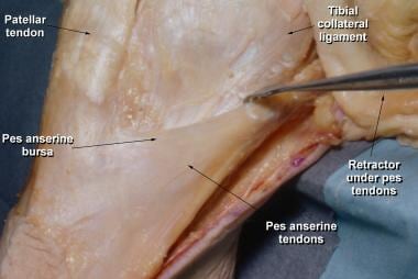 Pes Anserine Bursitis Background Anatomy Pathophysiology
 Flat Feet Eorthopod Com
 A B Schematic And Anatomical Representation Of Pes
 Flat Foot Pes Panus Anatomy Model
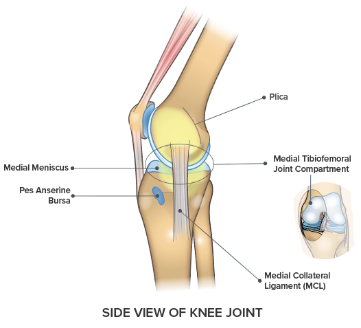 Knee Pain On The Inside Of Your Joint Causes Solutions
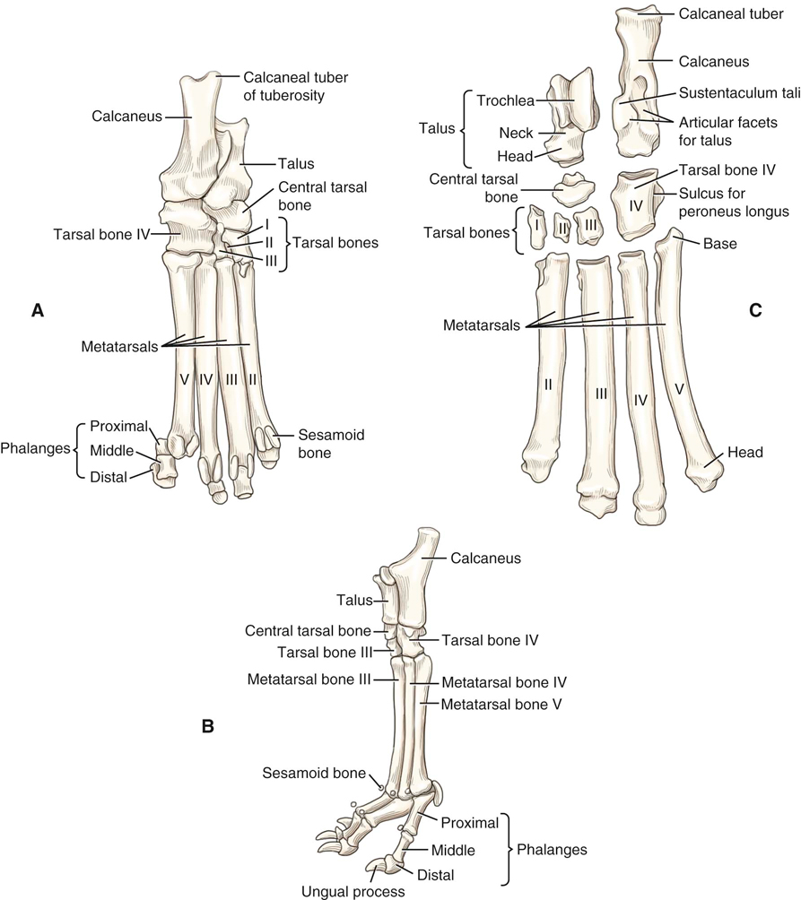 Canine Anatomy Veterian Key
 Anatomy Of A Rabbit Greeting Card
 Pes Anserinus Bursitis Symptoms And Treatment Bone And Spine
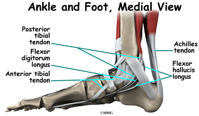
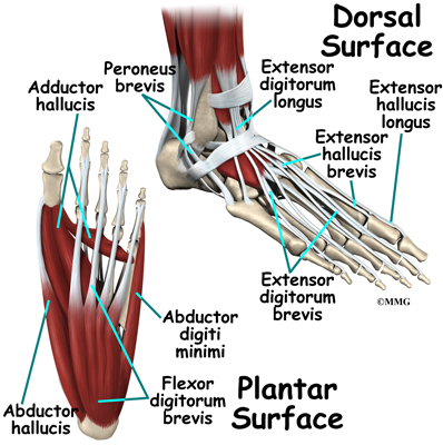
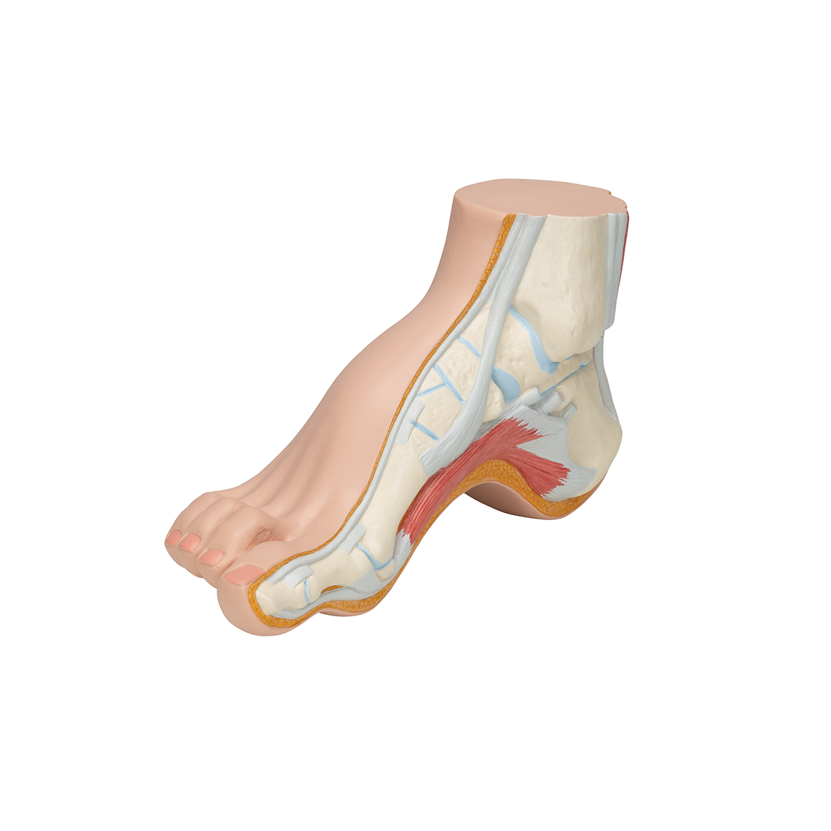
 Foot Wikipedia

 Pes Anserine Bursa Kneeguru
 Glossary Of Dinosaur Anatomy Wikipedia
 Anatomical Teaching Models Plastic Human Joint Models
 From Flat Foot To Fat Foot Structure Ontogeny Function
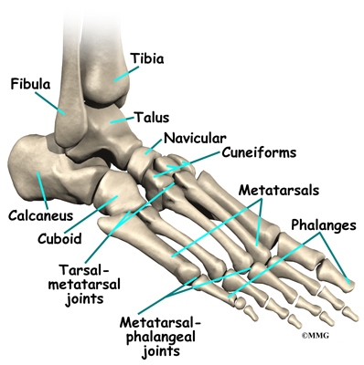
 Figure 5 Evolution Of Ankle Anatomy In Early Birds
 Canine Anatomy Veterian Key
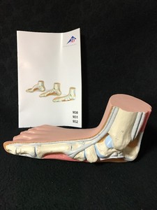 Details About 3b Scientific Flat Foot Pes Planus Anatomical Model Anatomy M31
Belum ada Komentar untuk "Pes Anatomy"
Posting Komentar