Shoulder Xray Anatomy
Knee shoulder shoulder arthrogram ankle elbow wrist hip contact. The extension of the shoulder series depends on the radiography department protocols and the clinical indications for imaging.

It is the most complete reference of human anatomy available on web ipad iphone and android devices.
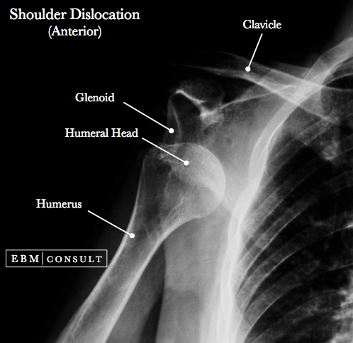
Shoulder xray anatomy. This effusion suggests intra articular fracture. This webpage presents the anatomical structures found on shoulder mri. The shoulder can dislocate posteriorly but anterior dislocation is approximately 50 times more common.
Anterior graphic of the shoulder. A plain x ray film of the shoulder may show dislocation osteoarthritis or a fracture of the humerus. X ray films cannot diagnose muscle or tendon injuries.
The shoulder series is fundamentally composed of two orthogonal views of the glenohumeral joint including the entire scapula. Quizzes about radiology anatomy quiz. E anatomy is an award winning interactive atlas of human anatomy.
Use the mouse to scroll or the arrows. Explore over 5400 anatomic structures and more than 375 000 translated medical labels. Ct mri radiographs anatomic diagrams and nuclear images.
Shoulder dislocation is a term often used loosely to indicate dislocation of the head of the humerus from the glenoid of the scapula. Shoulder radiographs are performed for a variety of indications including. Stanford bone tumor bayesian network issssr msk lectures for residents ocad msk cases from around the world stanford msk mri atlas has served almost 800000 pages to users in over 100 countries.
X ray shoulder x ray pelvis x ray hand pa ct head. Do not confuse with dislocation. The tendon of the subscapularis muscle attaches both to the lesser tubercle aswell as to the greater tubercle giving support to the long head of the biceps in the bicipital groove.
Click here to load quiz. Atlas of shoulder mri anatomy. An effusion or haemorrhage into the joint displaces the humeral head inferiorly.
Opening the quiz in incognito mode will prevent answers becoming pop up suggestions for future attempts. Usually secondary to trauma. Click on a link to get t1 axial view t2 fatsat axial view t1 coronal view t2 fatsat coronal view t2 fatsat sagittal view.
 The Shoulder
 Shoulder Imaging Shoulder Elbow Orthobullets
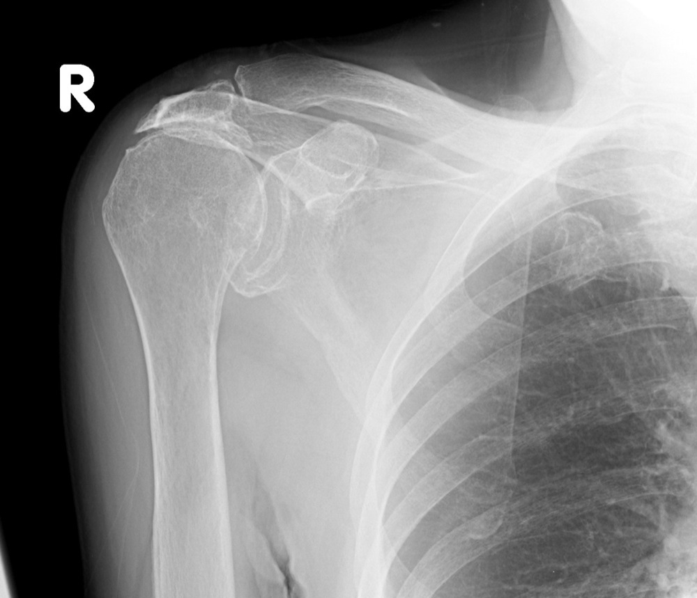
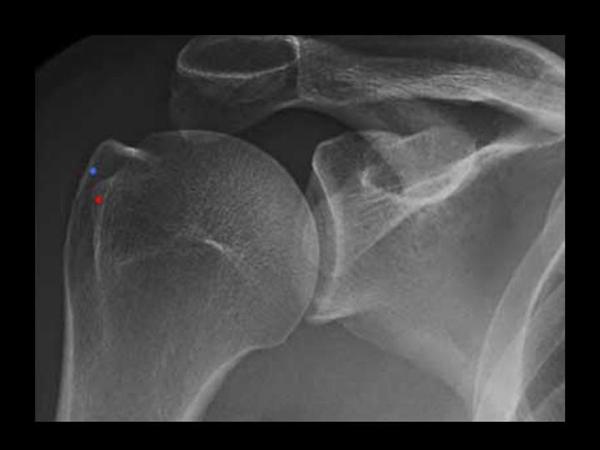
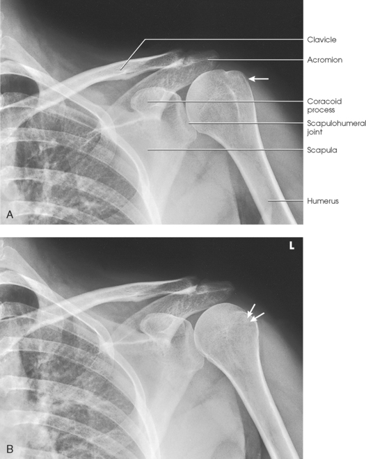
 Shoulder X Ray Labeled Anatomy Radiology Case
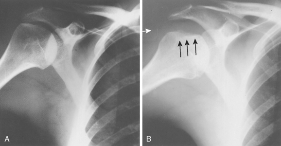
 Shoulder Arthritis Rotator Cuff Tears Causes Of Shoulder
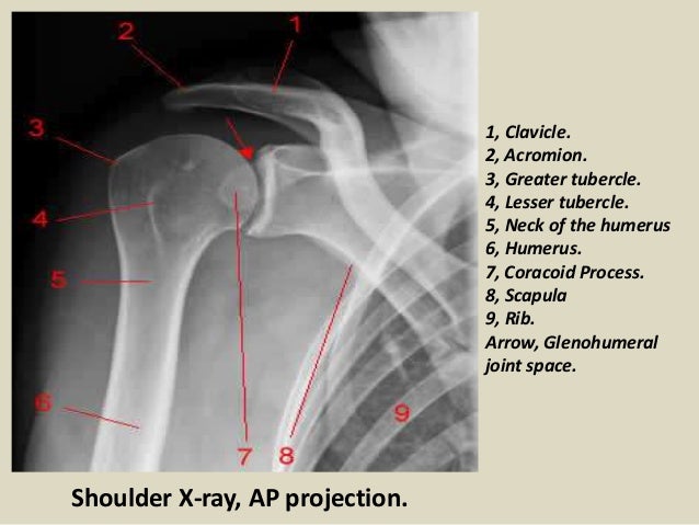
 Shoulder Anatomy And Normal Variants
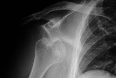
 Dislocated Shoulder Causes And Treatment The Hand Society
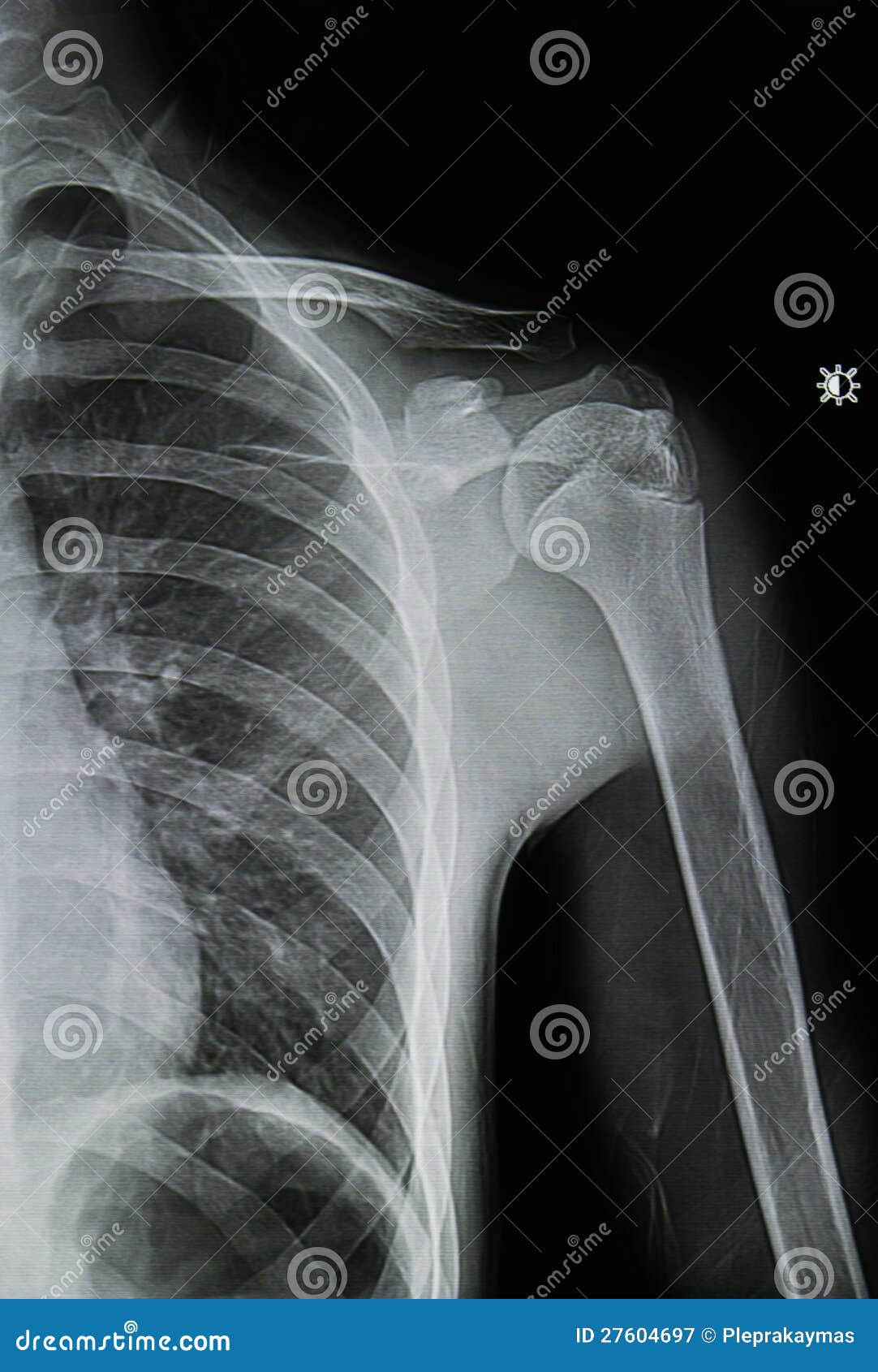 Shoulder X Rays Image Stock Image Image Of Body Bone
 Radiological Anatomy Of The Shoulder Arm Elbow Forearm
 Presentation1 Pptx Radiological Anatomy Of The Shoulder Joint
 Ap Shoulder External Rotation
 Shoulder Dislocation Practice Essentials Epidemiology
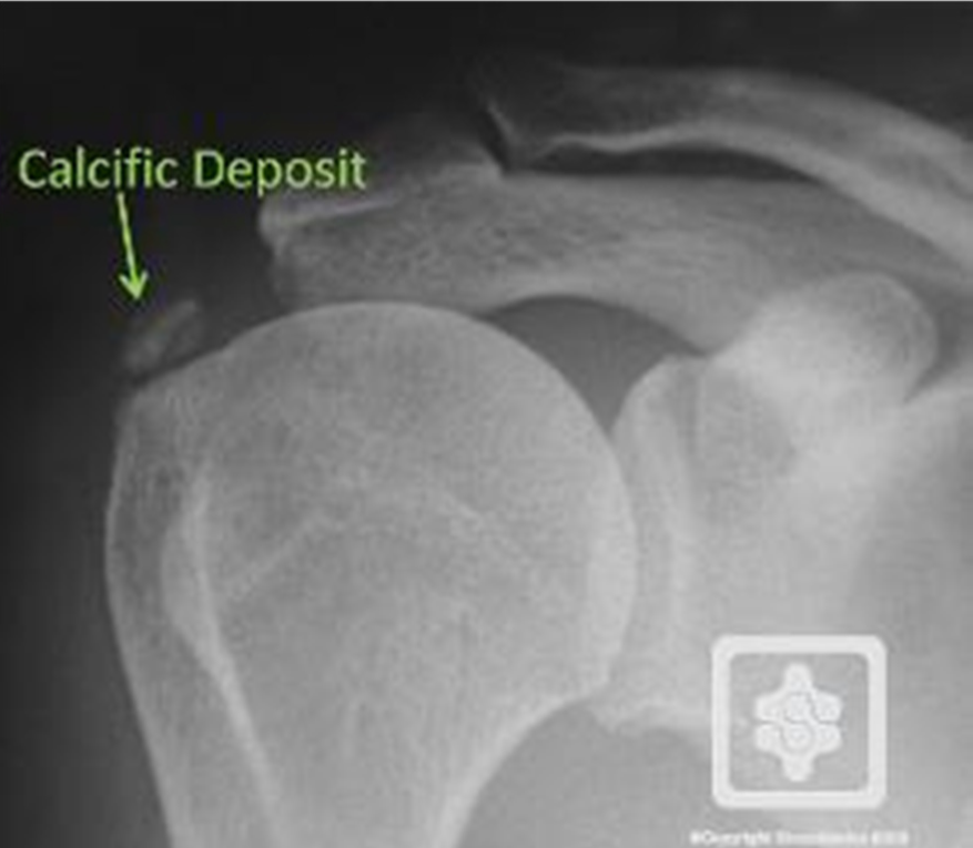
Calcific Tendonitis Brisbane Knee And Shoulder Clinic Dr
 Shoulder Radiology Springerlink
 Teaching Files University Of North Dakota
 Shoulder Girdle Radiology Key
 Radiographic Evaluation Of Shoulder Problems
:max_bytes(150000):strip_icc()/GettyImages-128118033-5b2713933418c60037a840a0.jpg) Proximal Humerus Bone Fractures Overview
Proximal Humerus Bone Fractures Overview
 Radiographic Evaluation Of Shoulder
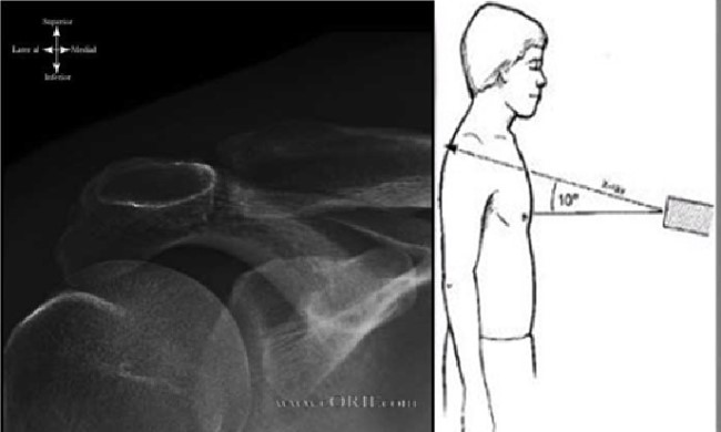
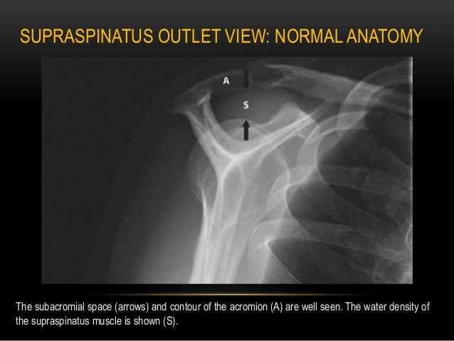
 Axillary View
 Zanca View Radiograph Demonstrating The Anatomy Of The

 Radiology Anatomy Images Surgical Neck Of Shoulder X Ray
 Shoulder Xray Images Stock Photos Vectors Shutterstock
 Ap Of The Shoulder Radiology Student Shoulder Anatomy

 Radiological Anatomy Of The Shoulder Arm Elbow Forearm
Belum ada Komentar untuk "Shoulder Xray Anatomy"
Posting Komentar