Spinal Cord Cross Section Anatomy
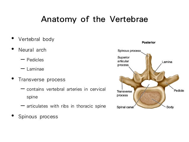
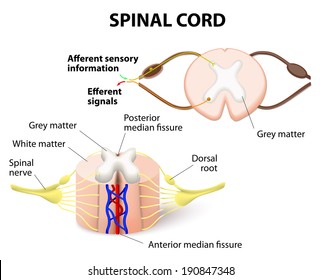
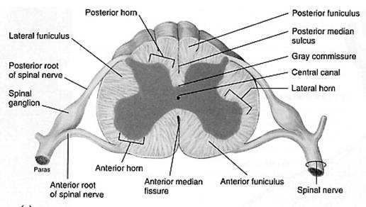
The ventral anterior median fissure and the more shallow dorsal posterior median sulcus. The central gray matter contains the neural cell bodies.
 Image Result For Spinal Cord Cross Sections Spinal Cord
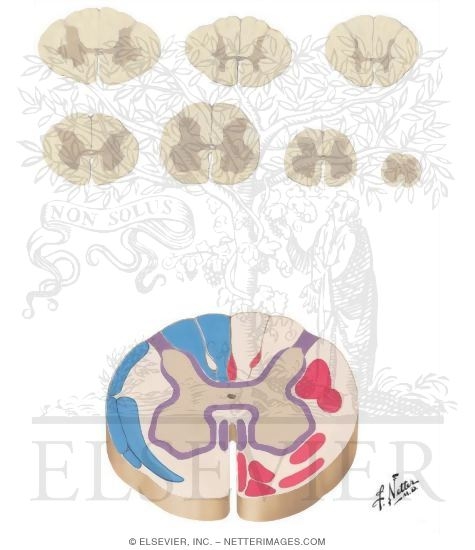
Cross sectional anatomy of the spinal cord the spinal cord appears to be somewhat flat with two grooves that mark its surface.

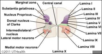
Spinal cord cross section anatomy. An interactive quiz covering spinal cord cross sectional anatomy through multiple choice questions and featuring the iconic gbs illustrations. Cross sectional anatomy of spinal cord the spinal cord like the brain consists of two kinds of nervous tissue called gray and white matter. Cross section of the spinal cord.
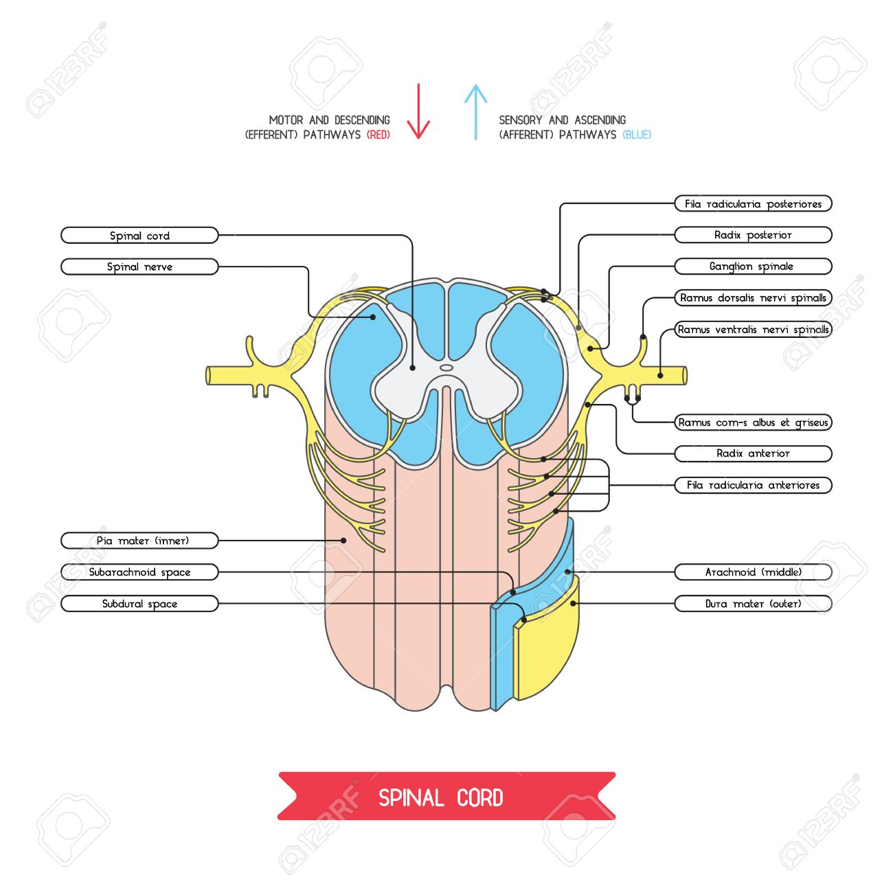
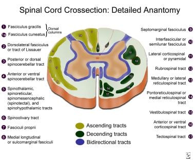
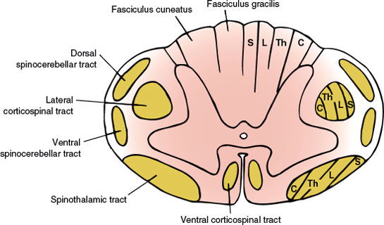
Cross section take a look at the spinal cord grey matter and fibers of the white matter tracts. The grey matter is butterfly shaped and surrounded by white matter. This article covers the anatomy of the spinal cord including its structure tracts and function.
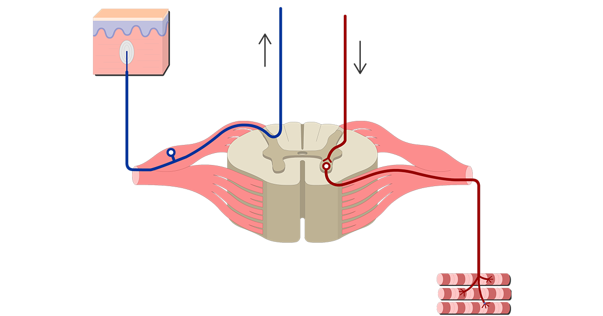
Sensory information is constantly sent to the brain while the motor information is sent to the muscles. Learn this topic now at kenhub. Neurological examination of the sensory system.
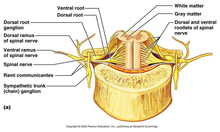
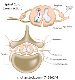
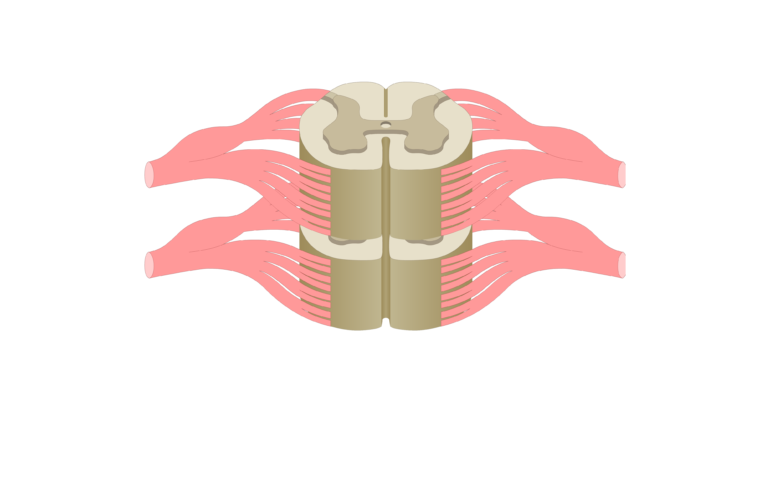
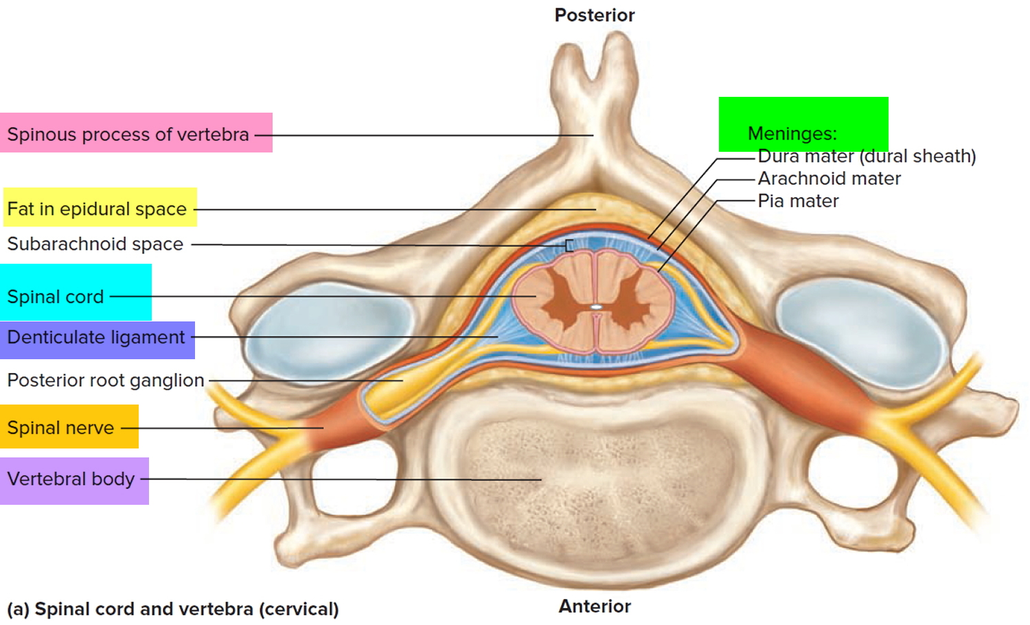
Learn vocabulary terms and more with flashcards games and other study tools. So we need some place from where the nerves can enter and exit the spinal cord right. A cross sectional view of the spinal cord demonstrates a central butterfly shaped area of gray matter and peripheral white matter fig.
The posterior median sulcus is the groove in the dorsal side and the anterior median fissure is the groove in the ventral side. Start studying spinal cord cross section anatomy. The spinal cord is elliptical in cross section being compressed dorsolaterally.
Two prominent grooves or sulci run along its length. When we observe the cross section we see the cord divided into grey matter and white matter. Gray matter has a relatively dull color because it contains little myelin.
The two grooves are named as follows. Internal anatomy of the spinal cord when viewed as a cross section from above the spinal cord consists of a butterfly shaped or thick h shaped region of gray matter that sits in the middle of the white matter. The central gray matter contains the neural cell bodies.
 Applied Cross Sectional Anatomy Of Spinal Cord
![]() Spinal Cord Anatomy Structure Tracts And Function Kenhub
Spinal Cord Anatomy Structure Tracts And Function Kenhub
 Cross Section Of Spinal Cord Central Nervous System Vector
 Neuroanatomy Online Lab 4 External And Internal Anatomy
 Cross Sectional Anatomy The Central Nervous System
 The Spinal Cord
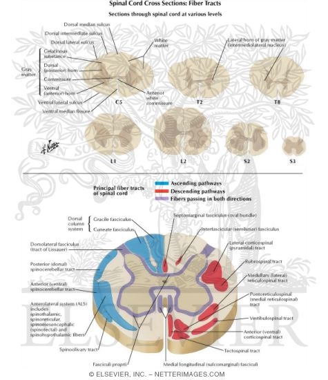 Sections Through Spinal Cord At Various Levels Spinal Cord
 Spinal Cord Anatomy Spine Orthobullets
 Spinal Cord Cross Section Images Stock Photos Vectors
 Anatomy Of Human Spinal Cord Both Cross Section And
 What Is The Spinal Cord What Is Its Anatomy And Function
 Cross Section Of Spinal Cord 1 Anatomy
 Spinal Cord Cross Section Diagram Spinal Cord Cross Section
 Spinal Nerve Roots
 Spinal Cord Cross Section Images Stock Photos Vectors
 Spinal Cord Cross Section Labeled Study Guide Anatomy
 Spinal Cord Cross Section Diagram A Cross Section Of The
 Cross Sectional Anatomy The Central Nervous System
 Topographic And Functional Anatomy Of The Spinal Cord Gross
 Spinal Cord Segments Cross Sectional Anatomy
 Anatomy Of The Spinal Cord
 Image Result For Spinal Cord Cross Sections Spinal Cord
 Sections Through Spinal Cord At Various Levels Spinal Cord
 Spinal Cord Anatomy Parts And Spinal Cord Functions
The Spinal Cord Human Anatomy And Physiology Lab Bsb 141
 Spinal Cord Neupsy Key
 Ch 12 Internal Anatomy Of The Spinal Cord
 Spinal Cord And Autonomic Ns
 Spinal Cord Anatomy Figure 12 6 A Cross Section Of The
 Ch 12 Gross Anatomy Of The Spinal Cord
 Cross Section Of Spinal Cord
 Applied Cross Sectional Anatomy Of Spinal Cord
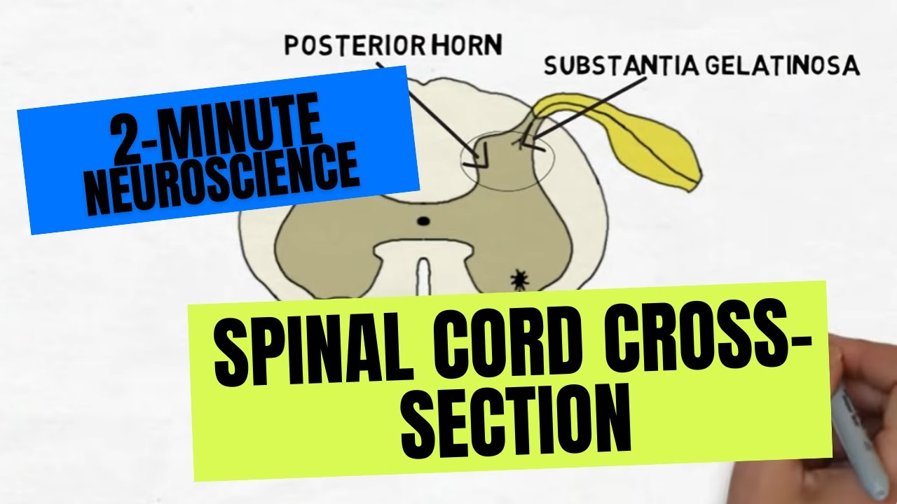 2 Minute Neuroscience Spinal Cord Cross Section
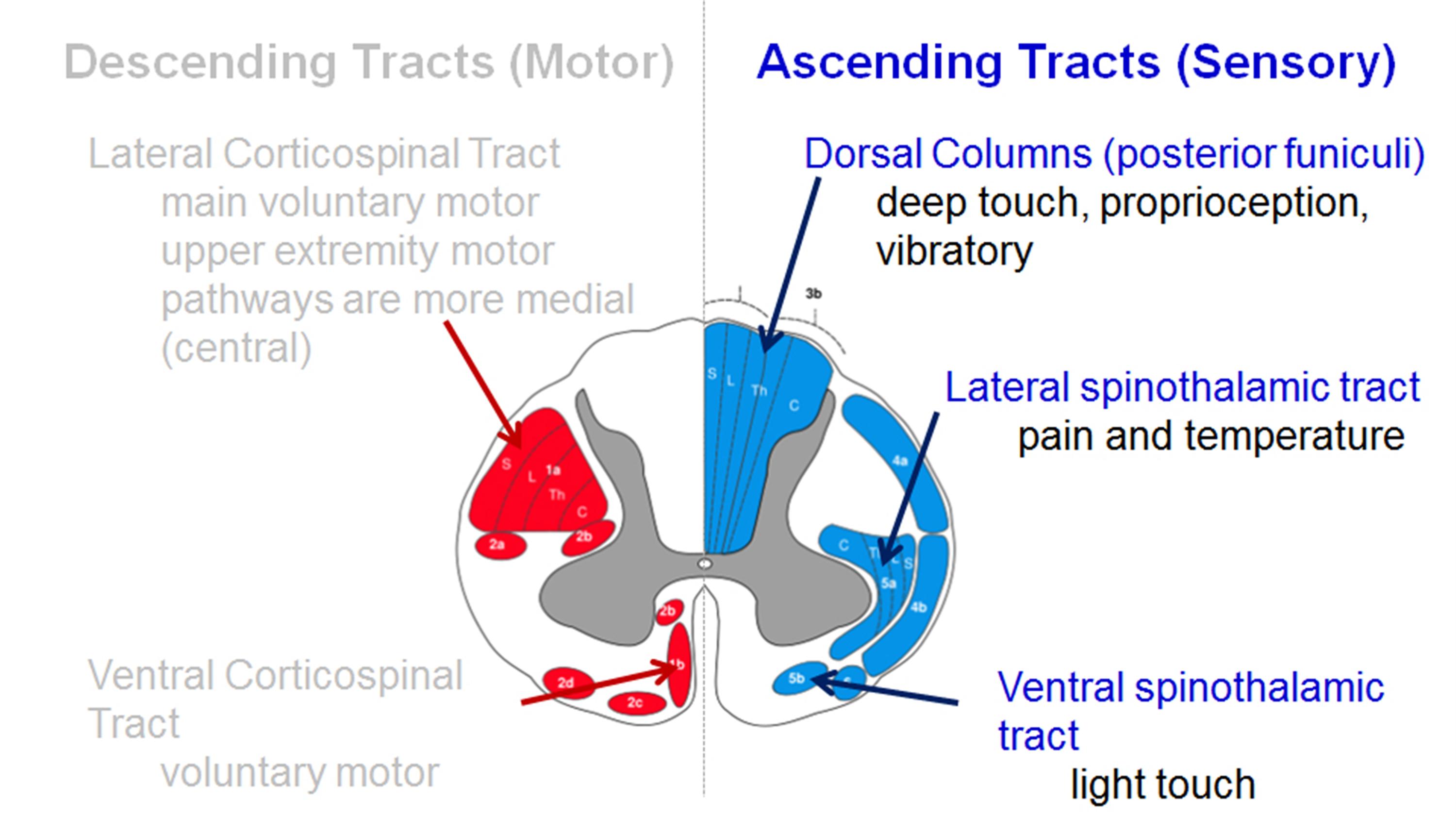
![]() Spinal Cord Ascending And Descending Tracts Kenhub
Spinal Cord Ascending And Descending Tracts Kenhub


Belum ada Komentar untuk "Spinal Cord Cross Section Anatomy"
Posting Komentar