Anatomy Of The Visual System
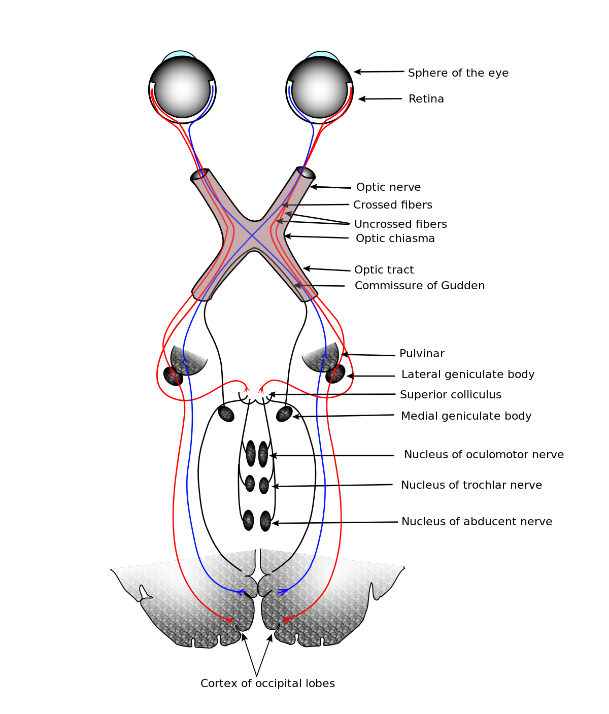
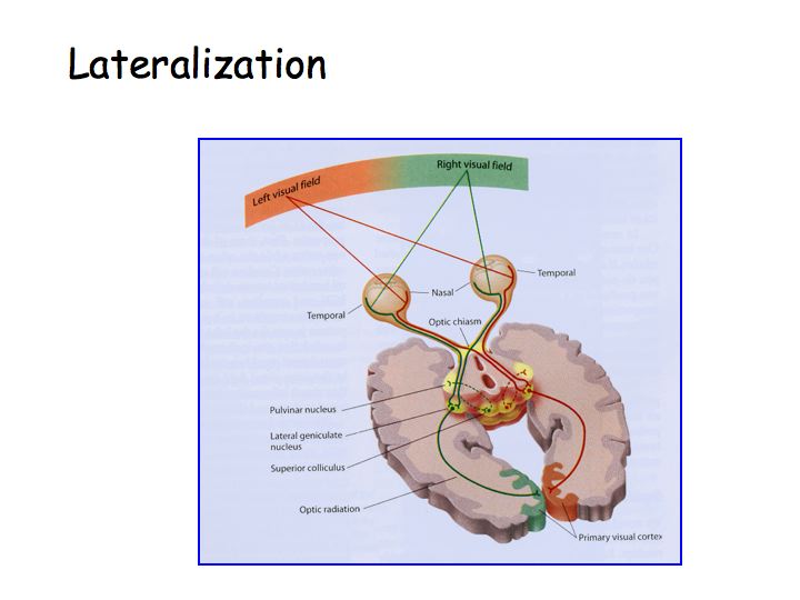
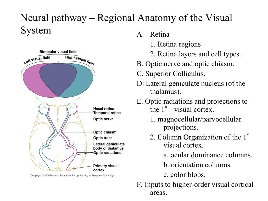
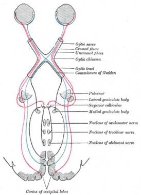
The inputs from each eye are combined at the optic chiasm and travel to the left lateral geniculate nucleus lgn of the thalamus then to the primary visual cortex also called v1 visual area 1 striate cortex or brodmann area 17 of the left hemisphere. The visual pathway to the occipital lobe of the brain consists most simply of a chain of five neurons.
 The Visual System Neuroanatomy Text And Atlas 4e
Comprehensive physiology coverage clarifies the integration between structure and function eliminating your need for multiple books on the anatomy and physiology of the visual system.

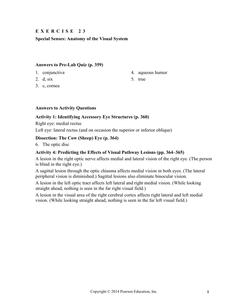
Anatomy of the visual system. 21 anatomy and physiology of the visual system. This concise source for the clinical anatomy of the visual system covers the clinical anatomy of the eye its adnexa and the visual pathways in a well illustrated well referenced format. 23 e x e r c i s e name lab timedate special senses.
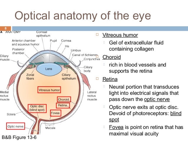
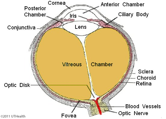
The iris is composed primarily of two smooth muscle layers one arranged radially and other circularly. The visual system carries out a number of complex tasks including the reception of light and the formation of monocular representations. It detects and interprets information from visible light to build a representation of the surrounding environment.
The visual system is the part of the central nervous system which gives organisms the ability to process visual detail as sight as well as enabling the formation of several non image photo response functions. The visual pathway to the occipital lobe of the brain consists most simply of a chain of five cells. Beginning with the pho toreceptor cell of the retina name them and note their location in the pathway.
Beginning with the photoreceptor cell of the retina name them and note their location in the pathway. Visual cortex of the cerebral hemisphere where is the lesion of someone with normal vision in left eye visual field. It balances histologic content of the microscopic anatomy with functional aspects of the eye and visual system.
The buildup of a nuclear binocular pe. Anatomyreview sheetof the visual system anatomy of the eye 1. Name five accessory eye structures that contribute to the formation of tears andor aid in lubrication of the eyeball and then name the major secretory product of each.
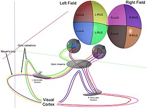
An emphasis on clinical application helps you better understand the processes that occur in disease and dysfunction. This textbook provides a comprehensive approach to the anatomy of the visual system and covers the embryology anatomy histology blood supply and innervation of the globe and ocular adnexa. Absence of vision in the right eye visual field right optic nerve.
 Special Senses Anatomy Of The Visual System Pages 1 6

 Visual System Wikipedia
 Visual System Wikipedia
 Ppt Vision Powerpoint Presentation Free Download Id 2114456
 Exercise 23 Anatomy Of The Visual System Diagram Quizlet
 Exercise Anatomyphysiologyrusso
 Special Senses Anatomy Of The Visual System Pages 1 6
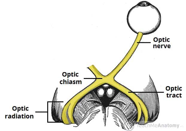 The Optic Nerve Visual Pathway Chiasm Tract
 Visual System Structure And Function
 Eye Conditions Retina Eye Consultants

 Understanding The Anatomy Of The Visual System A D A M Ondemand
 Pediagenosis
 The Visual System
 Perception Lecture Notes Lgn And V1
Exercise 23 Prt1 E X E R C I S E 2 3 Specialsenses
 Neuroanatomy Online Lab 7 Visual System Gross Anatomy
 Neuroanatomy Online Lab 7 Visual System Microscopic
%2C445%2C291%2C400%2C400%2Carial%2C12%2C4%2C0%2C0%2C5_SCLZZZZZZZ_.jpg) Clinical Anatomy Of The Visual System E Book Kindle
Clinical Anatomy Of The Visual System E Book Kindle
 Modern Neuro Ophthalmology Anatomy Physiology Of The
 Ppt Visual System Powerpoint Presentation Free Download
 Visual System Anatomy Overview Gross Anatomy
 Exercise 23 Special Senses Anatomy Of The Visual System
 Visual System Wikipedia
 Patient Education Visual System Nexj Health

 The Eyebody Patterns Eyebody
Belum ada Komentar untuk "Anatomy Of The Visual System"
Posting Komentar