Compact Bone Anatomy
Morris velilla 52995 views. Osteons are like thick tubes all going the same direction inside the bone similar to a bundle of straws with blood vessels veins and nerves in the center.
Cortical and cancellous bone.
Compact bone anatomy. Compact bone is dense so that it can withstand compressive forces while spongy cancellous bone has open spaces and supports shifts in weight distribution. Compact bone also called cortical bone dense bone in which the bony matrix is solidly filled with organic ground substance and inorganic salts leaving only tiny spaces lacunae that contain the osteocytes or bone cells. Together a canal and the osteocytes that surround it are called osteons.
Between the rings of matrix the bone cells osteocytes are located in spaces called lacunae. These cells are lined up in rings around the canals. Cortical bone is compact bone while cancellous bone is trabecular and spongy bone.
Resists stresses produced by weight and movement. Compact bone also known as cortical bone is a denser material used to create much of the hard structure of the skeleton. Video number anatomy 1 41.
Microscopic structures of compact bone wedge of bone duration. Overall the bones of the body are an organ made up of bone tissue bone marrow blood vessels epithelium and nerves. Provides protection and support.
Compact bone is made of special cells called osteocytes. The remainder is cancellous bone. Compact bone makes up 80 percent of the human skeleton.
Osteon or haversian system. Looks like solid hard layer of bone. It can be found under the periosteum and in the diaphyses of long bones where it provides support and protection.
Most of the skeleton is composed of this. In this tutorial we are going to examining the microscopic features that make up our long bone anatomy. Long bone compact bone and spongy bone duration.
Compact bone is the denser stronger of the two types of bone tissue. The osteon consists of a central canal called the osteonic haversian canal which is surrounded by concentric rings lamellae of matrix. Makes up the diaphysis of long bones and the external layer of all bones.
Small channels canaliculi radiate from the lacunae to the osteonic haversian. As seen in the image below compact bone forms the cortex or hard outer shell of most bones in the body. There are two types of bone tissue.
The basic structural and functional unit of compact bone opening in the center of an osteon carries blood vessels and layers of compact bone space containing osteocyte dimple looking things.
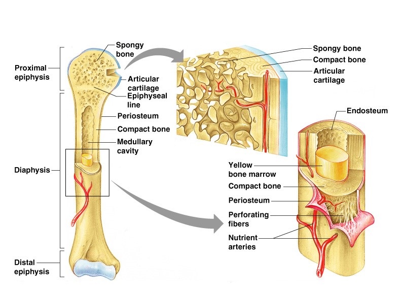 Structure And Functions Of Bones Online Science Notes
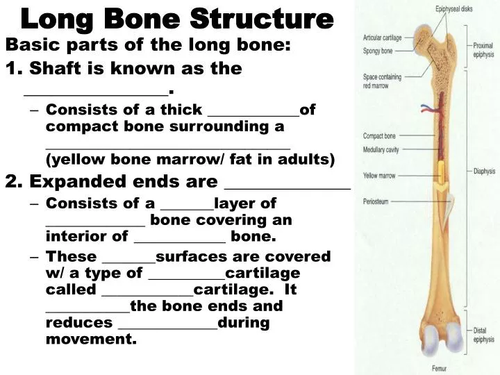 Ppt Long Bone Structure Powerpoint Presentation Free
 Structure Of Cortical Compact Bone
 33 2c Connective Tissues Bone Adipose And Blood
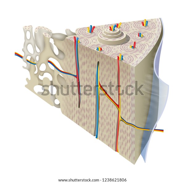 Anatomy Compact Bone Stock Illustration 1238621806
 Adv A P Ch 7 7 2 Microscopic Structure Of Bone
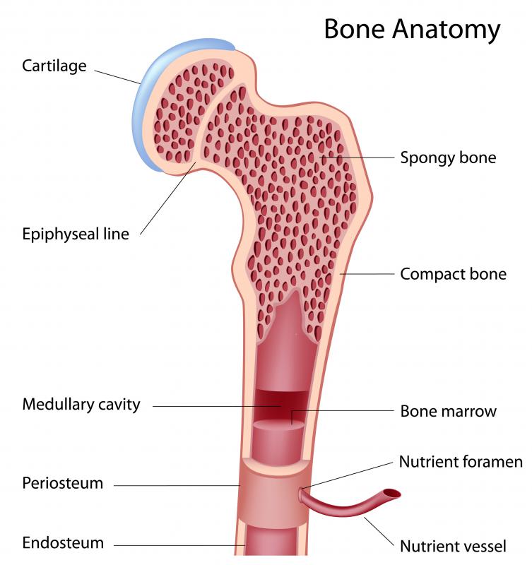 What Is The Function Of Compact Bone With Pictures
Microscopic Anatomy Of Bone Course Hero
 What Is Compact Bone Tissue Composed Of Socratic
 Anatomy Microscopic Structure Of Compact Bone Diagram
 Anatsc1102 Module 3 Compact Bone Structure Diagram Quizlet
 Structure And Function Of The Haversian System Explained
 Figure Anatomy Of The Bone The Pdq Cancer
Bone Structure And Function Simplified Dbriers Com
Compact Bone Spongy Bone And Other Bone Components Human
 Microscopic Structure Of Compact Bone Skeletal System
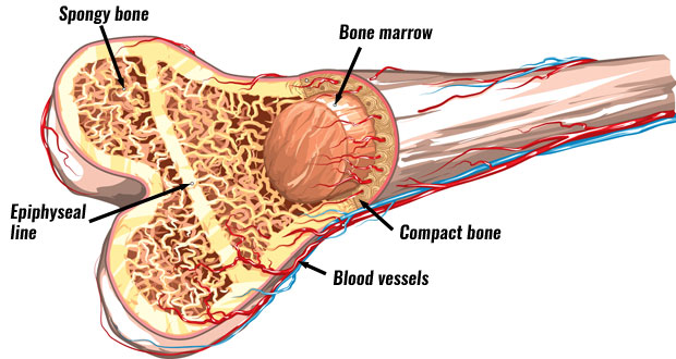 Bone Structure Anatomy Explained What Is Bone Marrow
 Structure Of Bones Biology For Majors Ii
 Structures Of Compact Bone Anatomy 213 With Woodman At
 14 4 Structure Of Bone Biology Libretexts
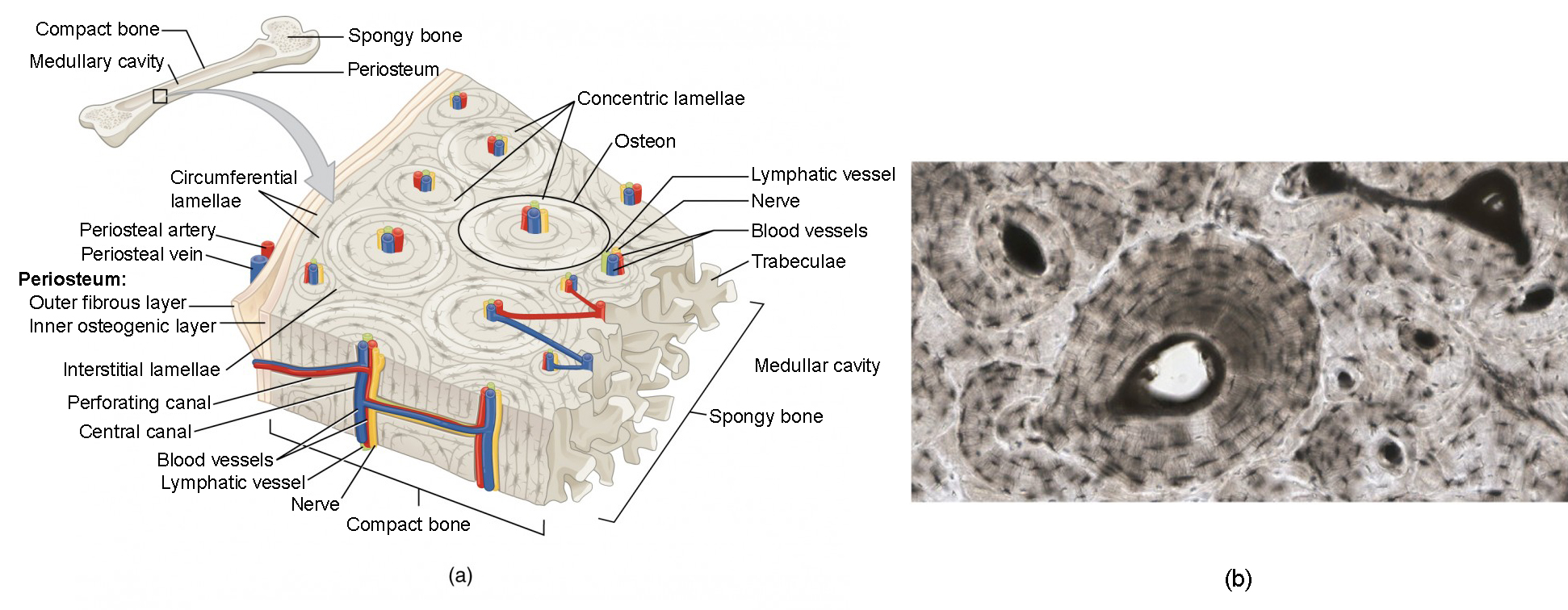 Bone Structure Anatomy And Physiology I


Belum ada Komentar untuk "Compact Bone Anatomy"
Posting Komentar