Microscopic Anatomy Of Skeletal Muscle
Returning the sarcolemma to its polarized state. What capability is most highly expressed in muscle tissue.
 Microscopic Anatomy Of Skeletal Muscle Flashcards Quizlet
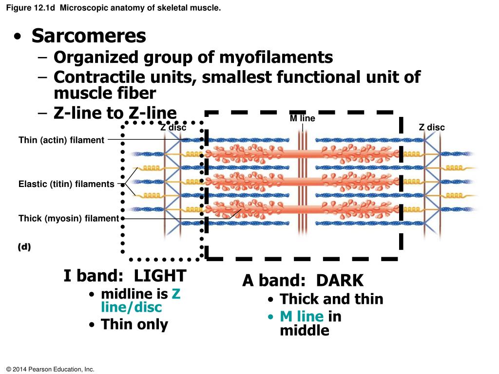
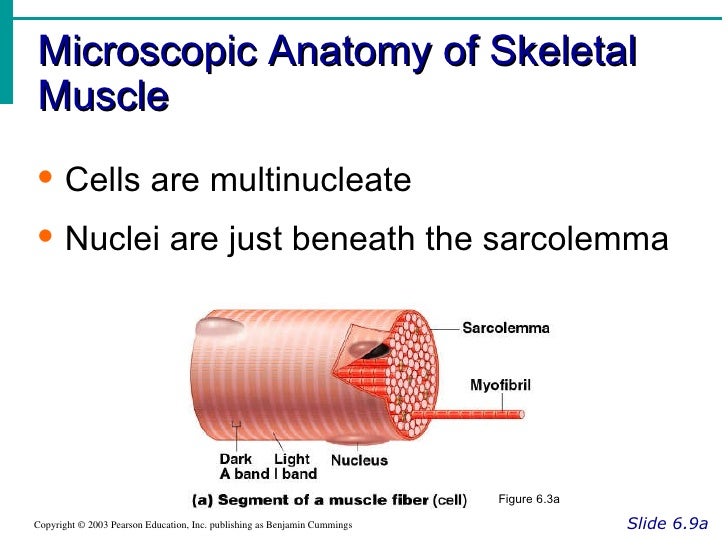
The microscopic anatomy of skeletal muscle have alternating light and dark bands.

Microscopic anatomy of skeletal muscle. Just pick an audience or yourself and itll end up in their incoming play queue. Microscopic anatomy of skeletal muscle. Microscopic anatomy and exercise14 organization of skeletal muscle review sheet 14 177 skeletal muscle cells and their packaging into muscles 1.
Microscopic anatomy of skeletal muscle learn with flashcards games and more for free. Microscopic anatomy and organization of skeletal muscle chapter 14. More channels open allowing more and more na into the cell.
Microscopic anatomy of skeletal muscle. Connective tissue ensheathing a bundle of muscle cells. Use the items on the right to correctly identify the structures described on the left.
Generation and propagation of an action potential. Generation of end plate potential. 0 0000 a shoutout is a way of letting people know of a game you want them to play.
Skeletal muscles are responsible for guarding the openings of the digestive and urinary tracts. The contractions of skeletal muscles pull on tendons and move elements of the skeleton. Ach binds opens na channels allowing it in.
As you know skeletal muscle tissue has alternating light and dark bands giving it a striated appearance. Microscopic anatomy of skeletal muscle. K channels open it rushes out at restores the negative charge inside.
Strengthen the muscle as a whole. Skeletal muscles support the weight of some internal organs. Skeletal muscles are responsible for the pumping action of the heart.
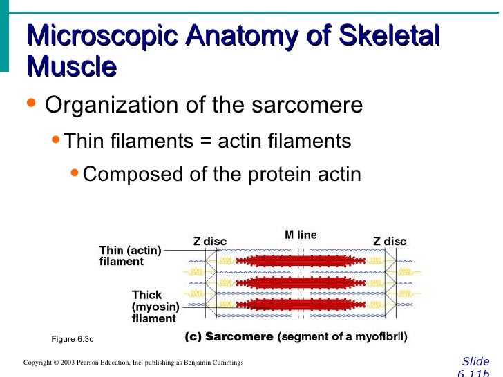
Light i dark a. Thin filaments composed of the contractile protein actin and some regulatory proteins that play a role in allowing or preventing myosin head binding to actin also called actin filaments are anchored to the z disk a disc like membrane light i band includes parts of two adjacent sarcomeres. Microscopic anatomy of skeletal muscle.
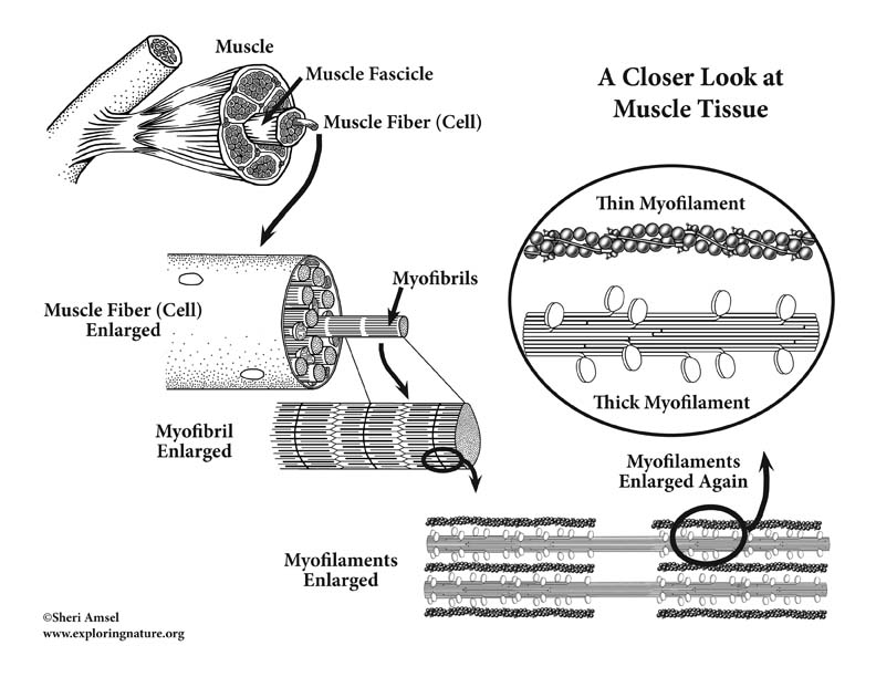
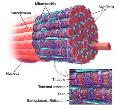
Plasma membrane that encloses the muscle cells contractile organelles found in the cytoplasm of muscle cells alternating bands along the length of the perfectly aligned my connective tissue surrounding a fascicle connective tissue ensheathing the entir gives the ability to move. Support and bind muscle fibres. Strengthen the connection of the muscle ego to the tendon.
Endomysium epimysium fascicle fiber myofilament myofibril perimysium sarcolemma sarcomere sarcoplasm and tendon. Provide a route for the entry and exit of nerves and blood vessels that serve the muscle fibres. The microscopic anatomy of skeletal muscle is made up of.
Microscopic anatomy and contraction of skeletal muscle we have already examined the structure of skeletal muscle as seen with the light microscope.
 Objective 3 Describe And Diagram The Microscopic Structure
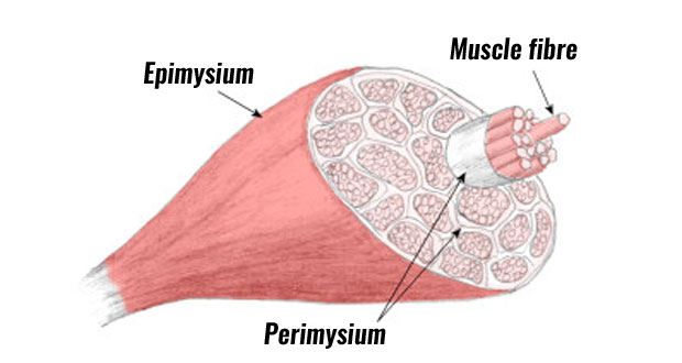 Muscle Anatomy Skeletal Muscle Structure Explained
 Microscopic A Amp P Muscle Webquest Microscopic Anatomy Of
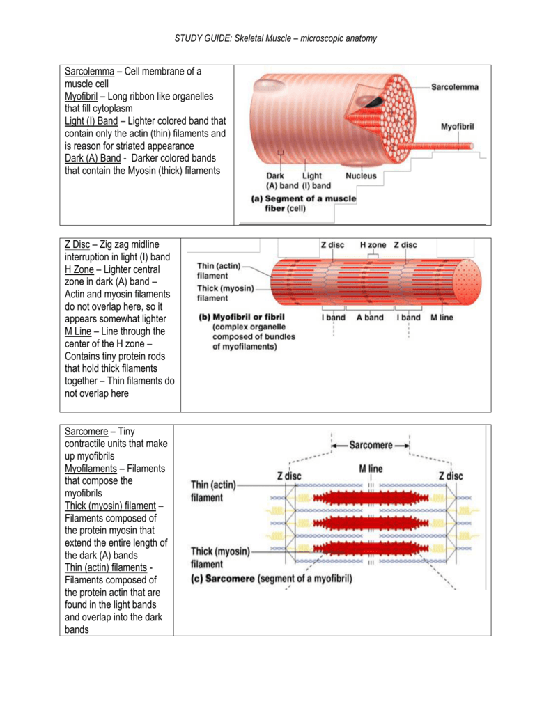 Study Guide Skeletal Muscle Microscopic Anatomy
 Macroscopic Microscopic Structure Of Muscular System
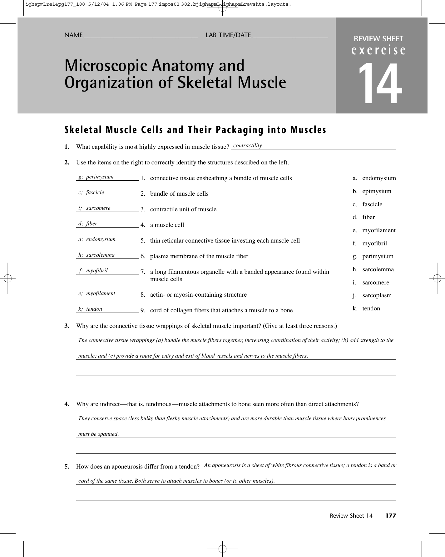 Exercise 14 Microscopic Anatomy And Organization Of Skeletal
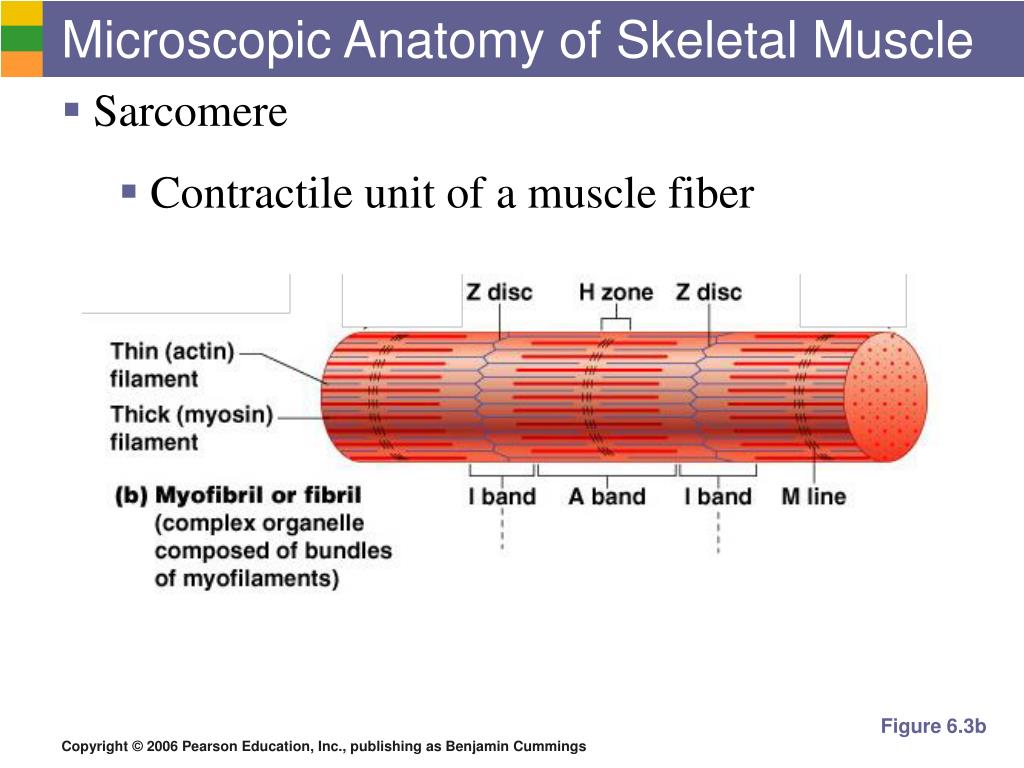 Ppt The Muscular System Powerpoint Presentation Free
 Ppt Exercise 14 Powerpoint Presentation Free Download
 Skeletal Muscle Handbook Of Microscopic Anatomy Vol 2 No
7 2 Microscopic Anatomy And Contraction Of Skeletal Muscle
 Microscopic Anatomy Of Skeletal Muscles Diagram Quizlet
 Muscular System Anatomy And Physiology Nurseslabs
 Muscle Contraction Microscopic Anatomy Of Skeletal Muscle
 Skeletal Muscle Microscopic Anatomy
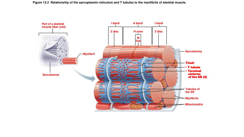 Figure 12 1 Microscopic Anatomy Of Skeletal Muscle Ppt
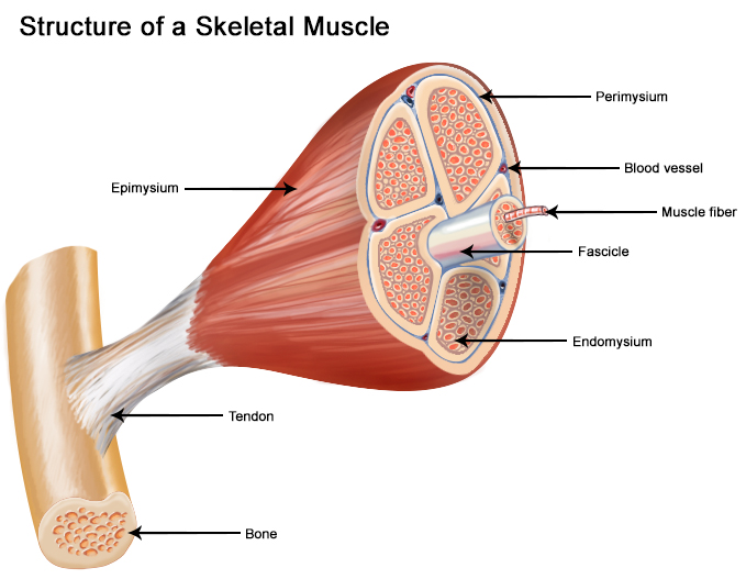 Seer Training Structure Of Skeletal Muscle
 Microscopic Anatomy Of Skeletal Muscle Youtube
Microscopic Anatomy Of Upper Arm Muscles Infobarrel Images
 Solved Microscopic Anatomy Of Skeletal Muscle Finst Id
 The Biomechanics Of Human Skeletal Muscle Basic
Belum ada Komentar untuk "Microscopic Anatomy Of Skeletal Muscle"
Posting Komentar