Equine Limb Anatomy
This order includes many species associated with livestock such as sheep goats pigs cows and camels as well as species of giraffes antelopes and deer. The distal limb bones are the foundation of equine lower leg.
 The Glass Horse Elements Of The Equine Distal Limb Vetbooks
Distal limb bones lower leg bones.
Equine limb anatomy. The sensitive connective tissue layer of the skin located below the epidermis containing nerve endings sweat glands sebaceous glands and blood. Equine lower limb anatomy. This is in contrast to even toed ungulates members of the order artiodactyla which walk on cloven hooves or two toes.
The fetlock joint is where the equine distal limb becomes more complex. Visit my other website anatomy of the equine to see more anatomy of the lower leg. 9 distal limb bones.
Equine distal limb we are not focusing on the distal limbdigit of the horse just because it is big and you can see everything. There are only three bones in this region. Select a body part and angle on the left then select the type of image from the top menu.
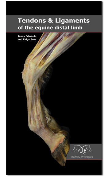
The content of the vea currently includes dissection of the equine head pelvic limb and thoracic limb with accompanying osteology of the limbs and skull. Equine distal limb tendons ligaments the success and handiness of our original hoof anatomy pocket guide led us to create this expanded in depth guide to the equine distal limb. In the young horse they are separate but often the splint bones fuse to the cannon bone.
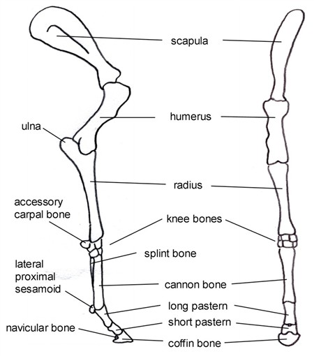
There are three bones between the knee and fetlock joints. Virtual equine anatomy vea is based on our first anatomy program virtual canine anatomy vca and is programmed with interactive elements including illustrative photographs of equine dissections. We are doing it because the equine digit is important to you and to the horse.
In the young horse there are three separate bones in the cannon region of the leg.
 Equine Distal Limb Muscles Pics Anatomy Gross Anatomy I
 Equine Limb Anatomy Puncture Wounds And Infections
 Novobrace Tendonitis Desmitis And Soft Tissue Injury
 Vitals Anatomy Horse Side Vet Guide
 Animals Free Full Text Measuring Volumetric Changes Of
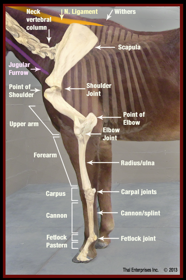
 Equine Limb Anatomy Horse Leg Anatomy Diagram Horse
 Hoofnotes Infographic Equine Anatomy Part 4 The
 Horse Anatomy Mobility Health
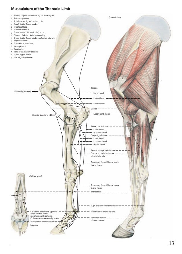
 The Musculature Of The Equine Thoracic Limb Anatomy Is
Vitals Anatomy Horse Side Vet Guide
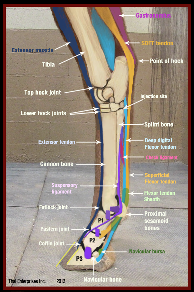
 Tendons Horse Anatomy Wikivet English
 Tendon Ligament Bone And Cartilage Anatomy Physiology
 Equine Limb Perfusion Illustrating The Hoof Puncture Wound
 Anatomy Of The Horse
 Nerve Distribution In The Metacarpus And Front Digit In The
 How Equine Forelimb Anatomy Plays Out With Conformation And
 Limbs Of The Horse Wikipedia
 Limb Vasculature Horse Anatomy Wikivet English
 The Equine Tarsus Hock Vet Physio Phyle
 Equine Limb Perfusion Illustrating The Hoof Puncture Wound
 Torn Horse Tendon The Long Road Back From This Equine
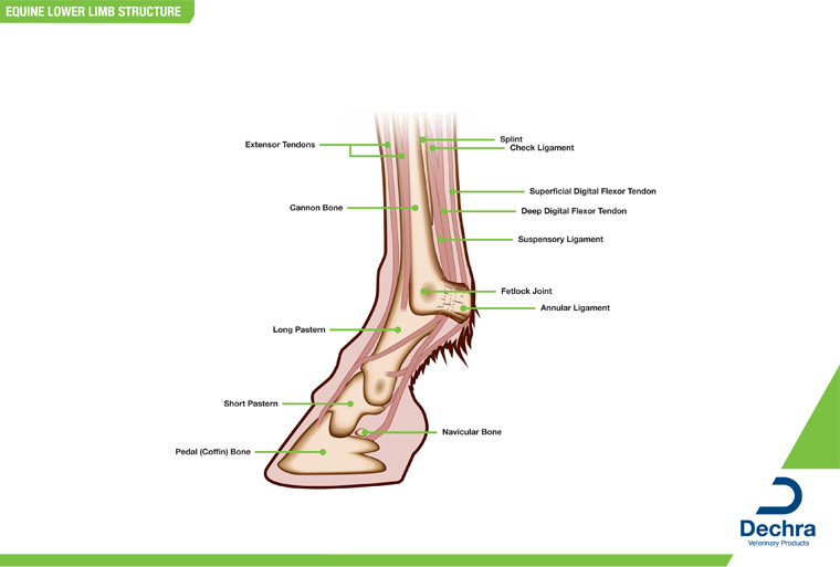 Downloads Anatomy Charts Dechra Veterinary Products
 Forelimb Muscle Activity During Equine Locomotion Journal
 Anatomy Of The Horse Osteology
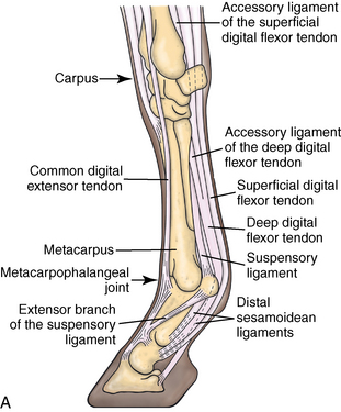
 Equine Distal Limb
 Novobrace Tendonitis Desmitis And Soft Tissue Injury
 The Effect Of Gait And Digital Flexor Muscle Activation On
 Fran Jurga S Hoof Blog News From Hoofcare Lameness
 Anatomy And Physiology Of Equine Tendons And Ligament
 Equine Distal Limb
 Equine Lower Limb Anatomical Horse Model Anatomy Alex Ridgeway
%2C445%2C291%2C400%2C400%2Carial%2C12%2C4%2C0%2C0%2C5_SCLZZZZZZZ_.jpg) The Equine Distal Limb An Atlas Of Clinical Anatomy And
The Equine Distal Limb An Atlas Of Clinical Anatomy And
 Fa16 Equine Medicine Surgery E1 Msk Cvm Musculoskeletal
 Equine Distal Limb Diagnostic Anaesthesia 1 Basic


Belum ada Komentar untuk "Equine Limb Anatomy"
Posting Komentar