Skull Anatomy Posterior
Posterior to the mandibular fossa on the external base of the skull is an elongated downward bony projection called the styloid process so named because of its resemblance to a stylus a pen or writing tool. Visit kenhub for more skeletal system quizzes.
 Cranial Vault Skull Base Diagnosis Skull Base Central
All vertebrates including humans have the same basic body plan they are strictly bilaterally symmetrical in early embryonic stages and largely bilaterally symmetrical in adulthood.
Skull anatomy posterior. Standard anatomical terms of location deal unambiguously with the anatomy of animals including humans. Quizzes on human skeletal system anatomy bone anatomy and bone markings. From anterior to posterior the fossae increase in depth.
Anatomy of the skull the skull is divided into the neurocranium or calvaria contains the brain and its meningeal coverings and the viscerocranium facial skeleton. In humans these sensory structures are part of the facial skeleton. The posterior and lateral views of the skull show us important bones that maintain the integrity of the skull.
Posterior skull bone markings quiz. Depression formed by squamous and mastoid temporal bone plus occipital bone. Visit kenhub for more skeletal system quizzes.
On the posterior skull the sagittal suture terminates by joining the lambdoid suture. Quizzes on human skeletal system anatomy bone anatomy and bone markings. The skull forms the anterior most portion of the skeleton and is a product of cephalisation housing the brain and several sensory structures such as the eyes ears nose and mouth.
That is they have mirror image left and right halves if divided down the middle. The lambdoid suture extends downward and laterally to either side away from its junction with the sagittal suture. The posterior surface protects the region of the brain that contains the occipital lobes and cerebellum.
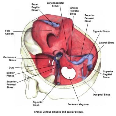
By teachmeseries ltd 2019 fig 13 lateral view of the skull showing the path of the meningeal arteries. The skull is composed of 22 bones excluding the middle ear ossicles with 8 forming the cranium and 14 forming the face. Inside the skull the base is subdivided into three large spaces called the anterior cranial fossa middle cranial fossa and posterior cranial fossa fossa trench or ditch link.
 Viewmedica Stock Art Skull Spinal Column And Rib Cage With
 Skull Anatomy Terminology Dr Barry L Eppley
 Frontal Bone Human Skull
 The Skull Summary Of Anatomy Docsity
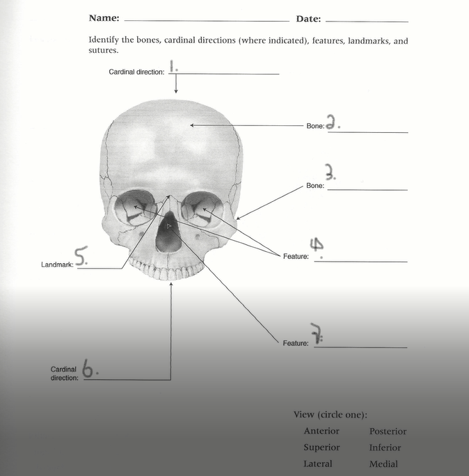 Solved Name Date Identify The Bones Cardinal Direction
 Human Being Anatomy Skeleton Child S Skull Image
 Posterior Skull Anatomy Diagram Quizlet
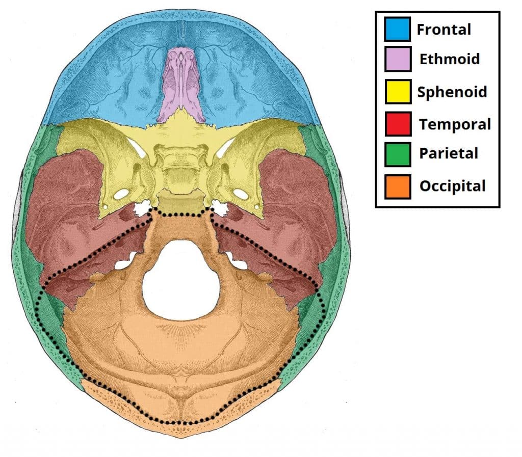
Posterior Cranial Fossa Boundaries Contents Teachmeanatomy
 Easy Notes On Skull Learn In Just 4 Minutes Earth S Lab
 Bones Of The Head Atlas Of Anatomy
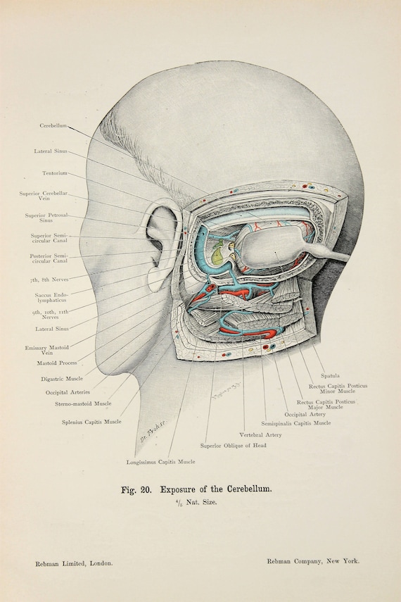 Skull Anatomy Cerebellum Ears Hearing C 1900 Double Sided Antique Anatomy Print Colour Anatomical Print Lithograph
 Human Skull Chart
 Structures Passing Trough Foramen Of Skull Foramen Of Skull
 Skull Anatomy Anterior And Posterior View
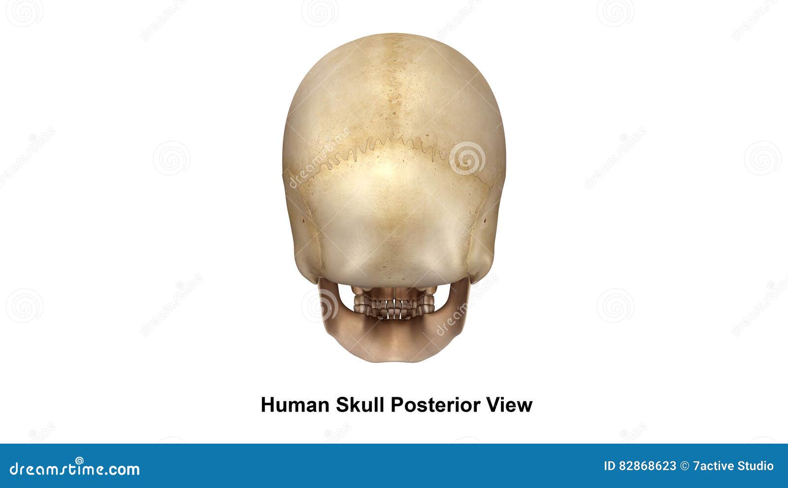
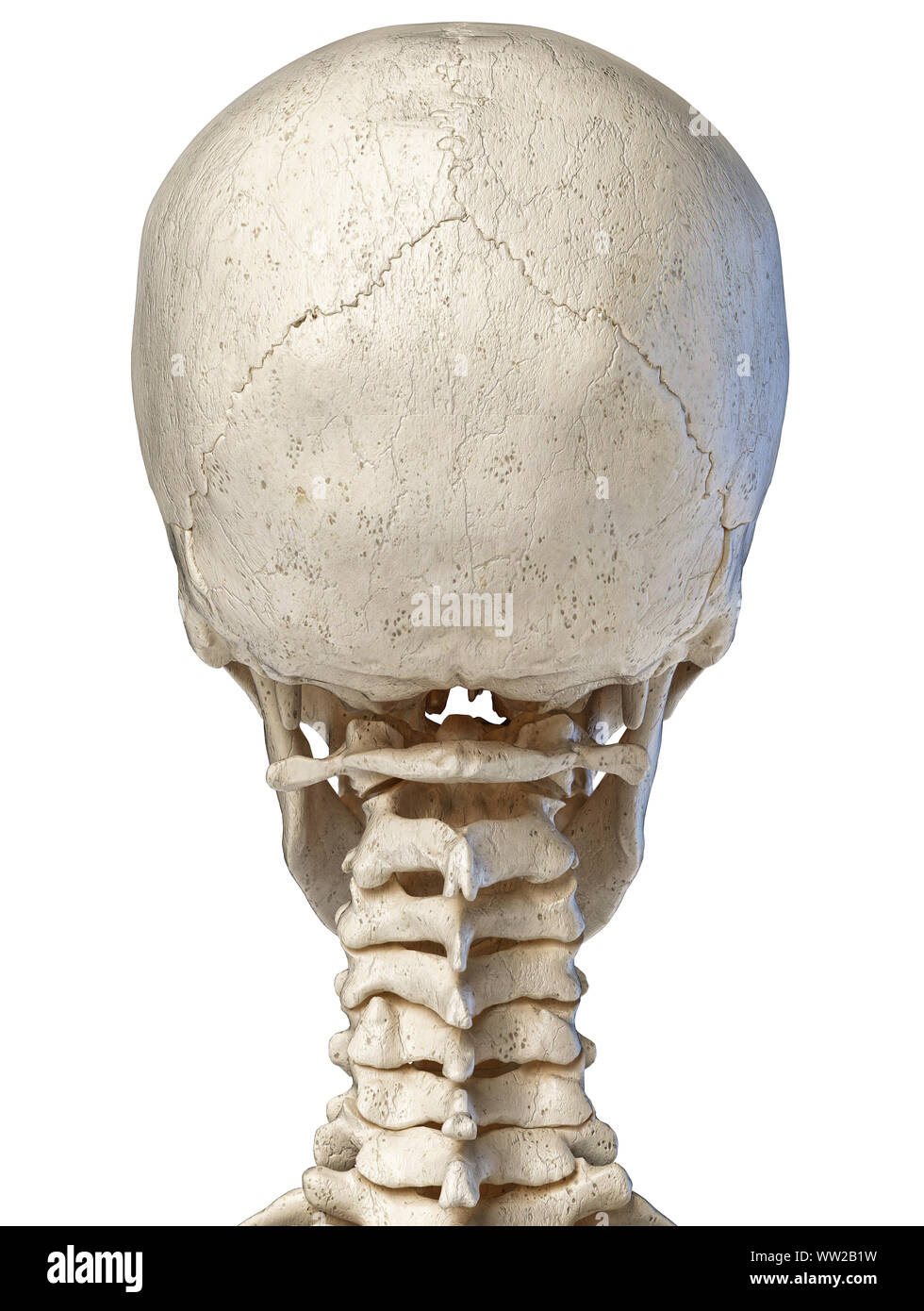 Anatomy Human Skull Rear View Stock Photos Anatomy Human
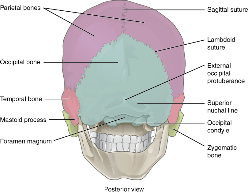 7 2 The Skull Anatomy And Physiology
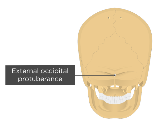 Occipital Bone Anatomy
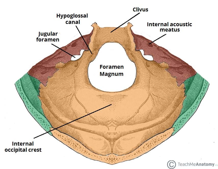 Posterior Cranial Fossa Boundaries Contents Teachmeanatomy
 Posterior Triangle Of The Neck Cervical Vertebrae Bone Head
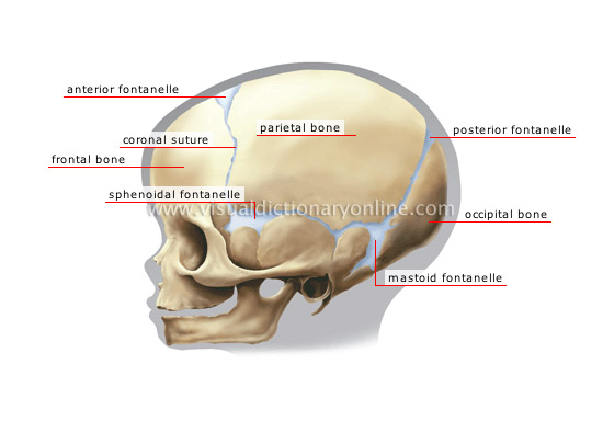
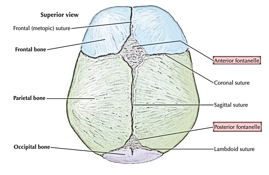
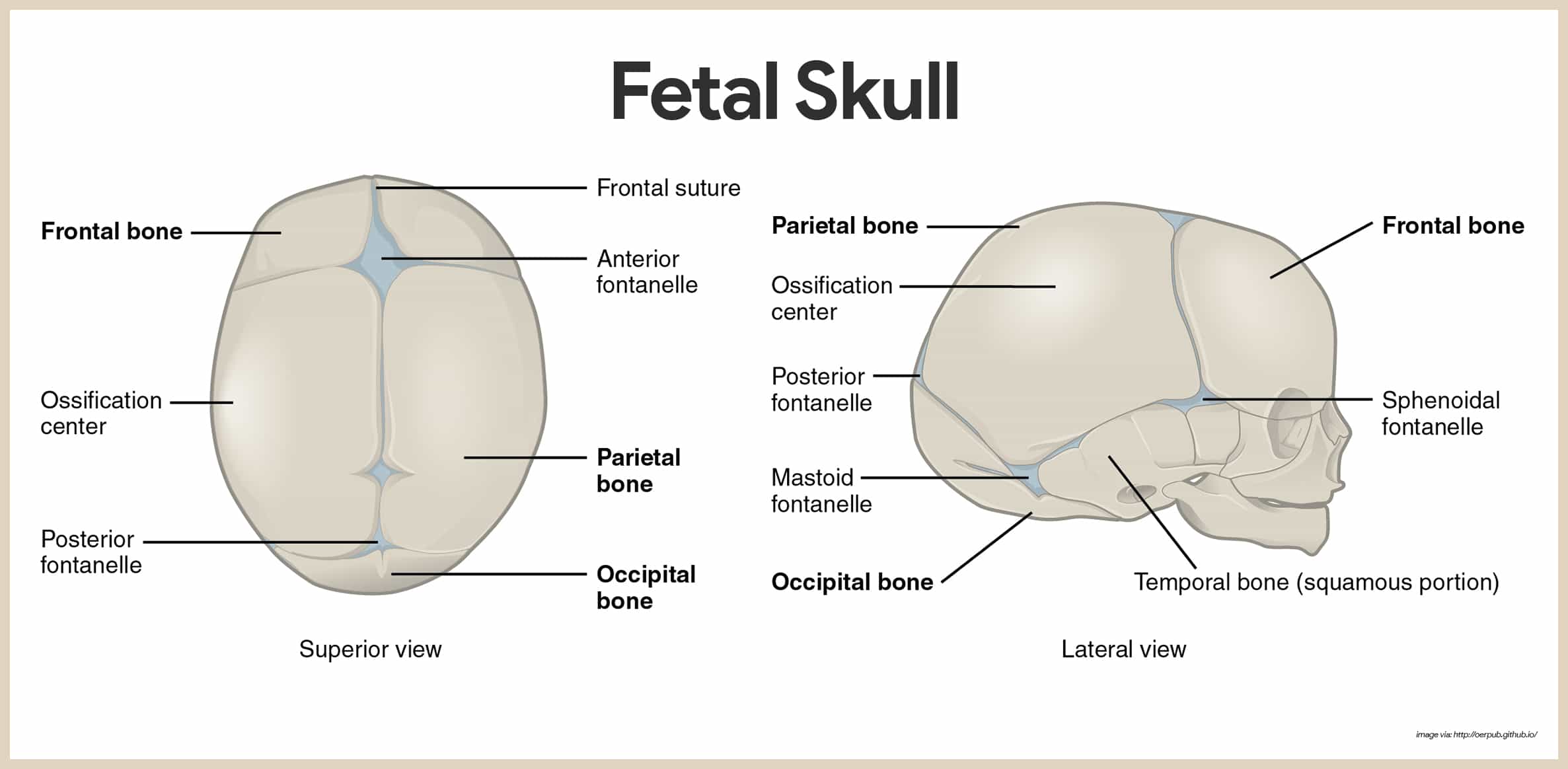
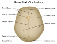 Anatomy Of The Newborn Skull
 Skull Base Anatomy Overview Anterior Skull Base Middle
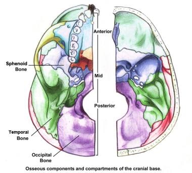 Skull Base Anatomy Overview Anterior Skull Base Middle
 Anatomy Of The Anterior Posterior Aspects Of Skull
 Skeletal System Anatomy And Physiology Nurseslabs
 Skull Posterior View Stock Illustration Illustration Of


Belum ada Komentar untuk "Skull Anatomy Posterior"
Posting Komentar