Ankle Anatomy Mri
This webpage presents the anatomical structures found on ankle mri. Mri anatomy of the ankle tendons and ligaments normal mri tendon anatomy tendons around the ankle are divided into four groups.
 The Radiology Assistant Ankle Mri Examination
This joint is a main contributor of stability in the lower limbs and it allows humans to perform actions such as running jumping and walking 1 2.

Ankle anatomy mri. It is also a fundamental communication tool to teach patients anatomy and pathology. The exquisite soft tissue contrast resolution noninvasive nature. Uq med year 1 gafradiographic anatomy lower limb.
Rmhalf msk ankle. 663 3 normal extremity. Stanford bone tumor bayesian network issssr msk lectures for residents ocad msk cases from around the world stanford msk mri atlas has served almost 800000 pages to users in over 100 countries.
It carries the weight of the body and can undergo a myriad of pathology most commonly traumatic injuries of the medial and lateral malleoli. Mri of the ankle. The tibia extends inferiorly to articulate with.
This module is a comprehensive and affordable learning tool for medical students and residents and especially for physicians anatomists rheumatologists orthopaedic surgeons and radiologists. Mri of the ankle and feet. Screen on fatsat images for bone marrow edema.
Scroll through the image stack for the. Once you have studied the bones scan the joints for effusion. Ankle mri examination systematic approach.
Magnetic resonance mr imaging has opened new horizons in the diagnosis and treatment of many musculoskeletal diseases of the ankle and foot. The ankle joint is comprised of the tibia fibula and talus as well as the supporting ligaments muscles and neurovascular bundles. It demonstrates abnormalities in the bones and soft tissues before they become evident at other imaging modalities.
Racsuq advanced surgical anatomy course upper and lower limbs. Knee shoulder shoulder arthrogram ankle elbow. Anterior posterior medial and lateral.
Click on a link to get sagittal view t1 axial view t2fatsat coronal view t2fatsat sagittal view t2fatsat. Start your exam with fatsat images of the bones to screen for edema. Use the mouse to scroll or the arrows.
Internal derrangements of joints. 37 magnetic resonance imaging mri the ankle is the joint that is located between the leg and the foot.
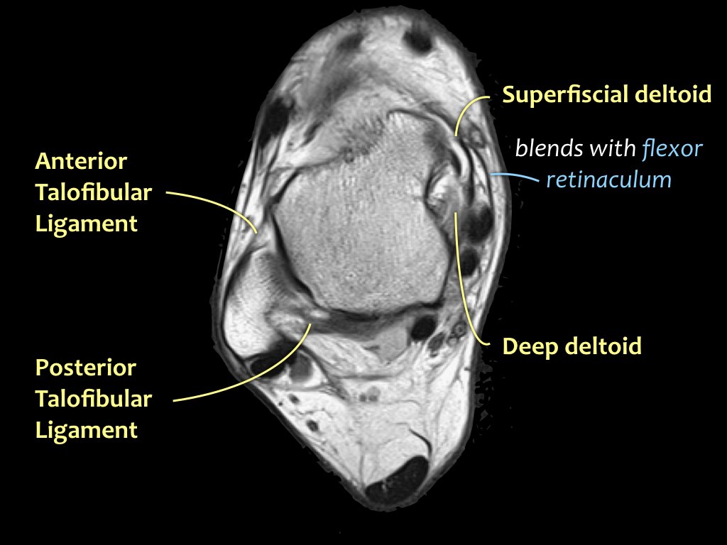
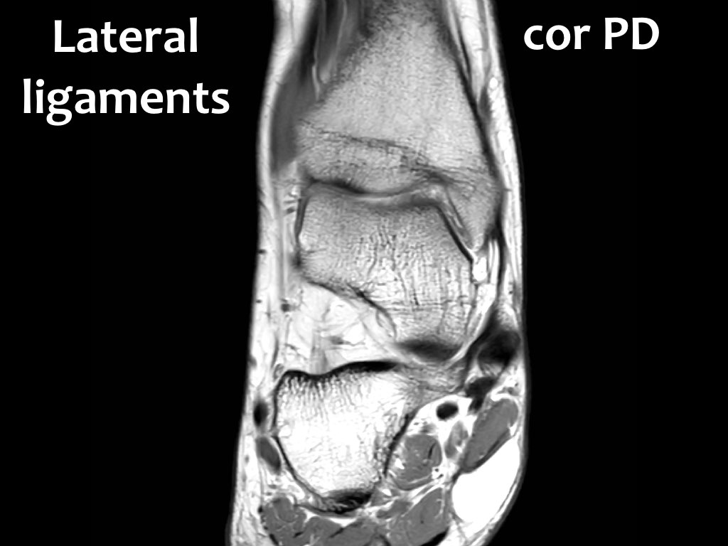
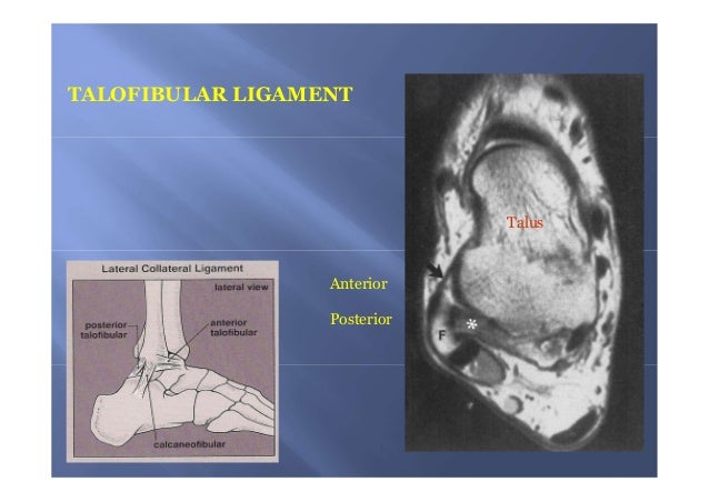
 Mri Anatomy Of Lateral Ankle Ligaments
 Mri Imaging Of Soft Tissue Tumours Of The Foot And Ankle
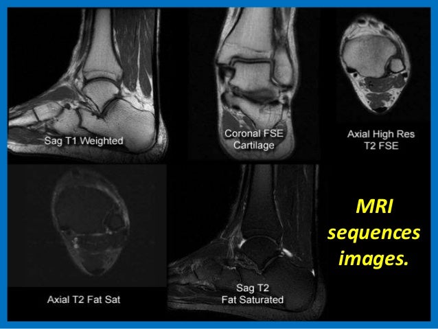
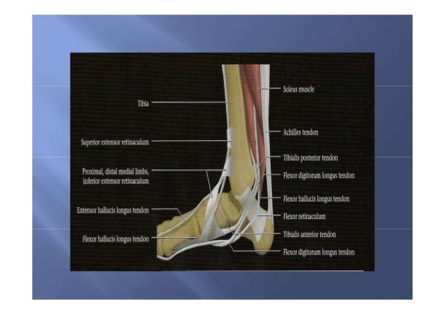
 Mri Of The Ankle Detailed Anatomy
 Aspetar Sports Medicine Journal Mri Of Ankle And Foot
 Magnetic Resonance Imaging Of Ankle Ligaments A Pictorial
 Causes Of Foot Heel Ankle Pain
 Pigmented Villonodular Synovitis Pathology Orthobullets
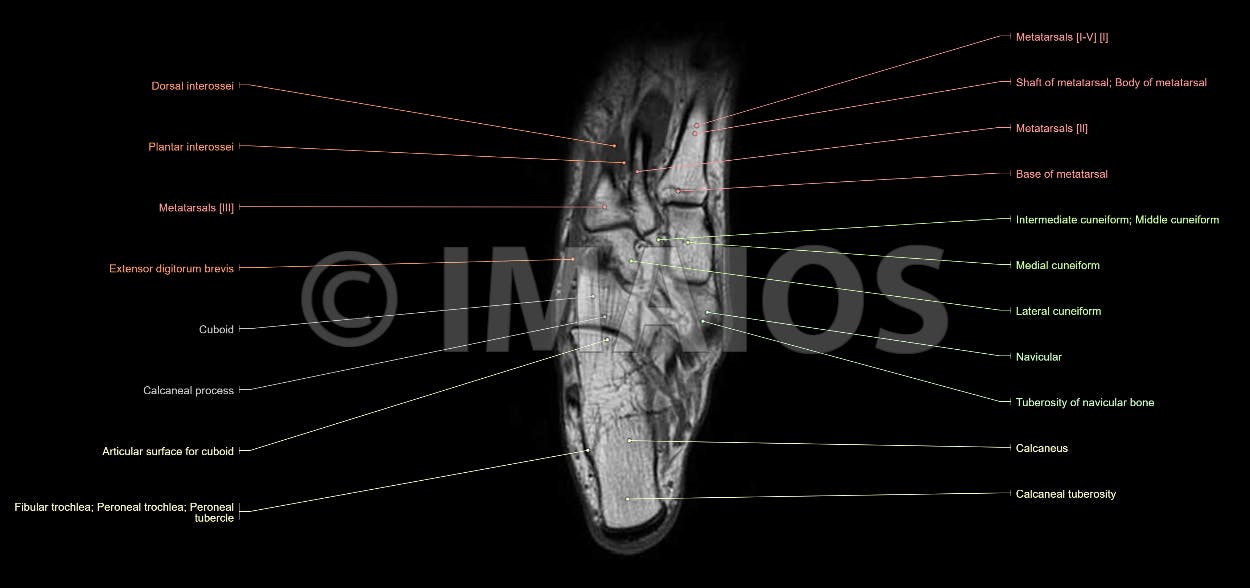 Anatomy Of The Foot And Ankle Mri
 Mri Lower Extremities Leg Cedars Sinai
 Presentation1 Pptx Ankle Joint
 Normal Ankle Mri Radiology Case Radiopaedia Org
 Courses Mri Online
 The Radiology Assistant Ankle Mri Examination
 Sinus Tarsi Syndrome Eurorad
 Stanford Msk Mri Atlas C 2019
 Mri Anatomy Of Ankle
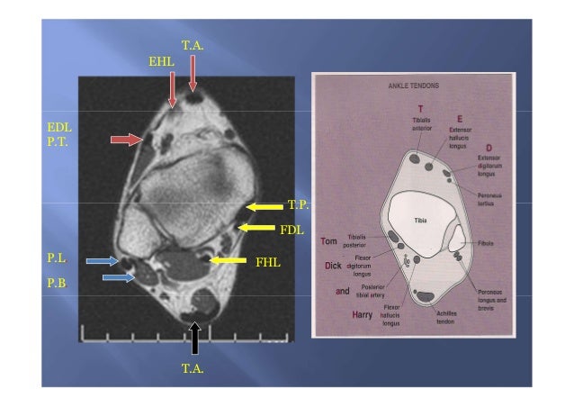
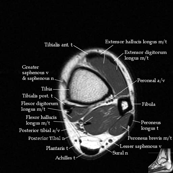
 Ankle Tendons Topographic Anatomy Radiology Case
 Mri Anatomy Of Ankle
 Mri Anatomy Of Ankle
 Ankle X Rays
 The Radiology Assistant Ankle Mri Examination
 Mri Of The Ankle Detailed Anatomy
 Mri Of The Ankle Detailed Anatomy
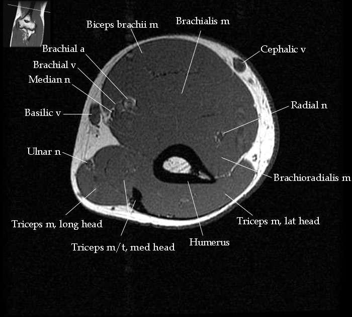 Mri Elbow Anatomy
 Anatomy Of The Foot And Ankle Mri
 Mri Anatomy Of Ankle
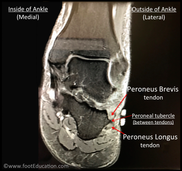 Peroneal Tendonitis Footeducation
 Mri Of The Ankle Detailed Anatomy
 Mri Ankle Anatomy


Belum ada Komentar untuk "Ankle Anatomy Mri"
Posting Komentar