External Nose Anatomy
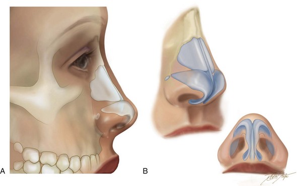
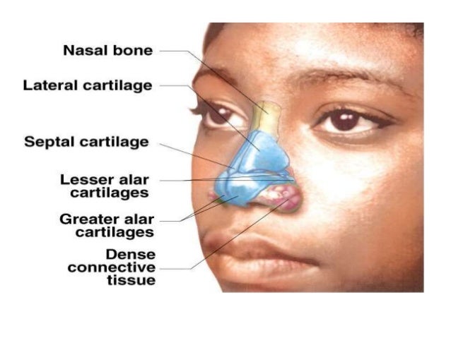
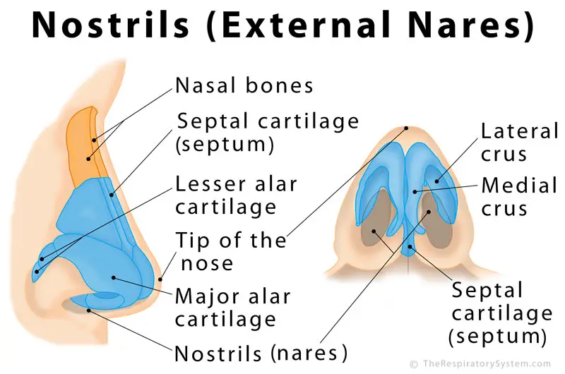
The lateral and major alar cartilages are the largest and contribute the most to the shape of the nose here. The nose is made up of.
 External Nose Anatomy Qa
Spanning between the root and apex is the dorsum of the nose.

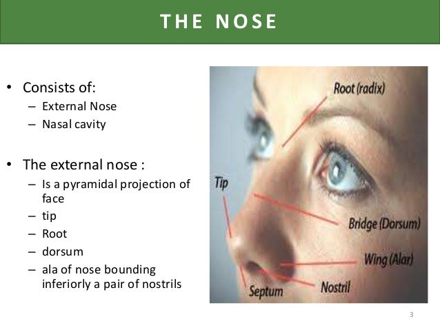
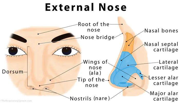
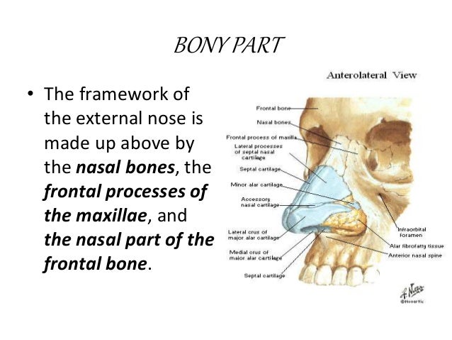
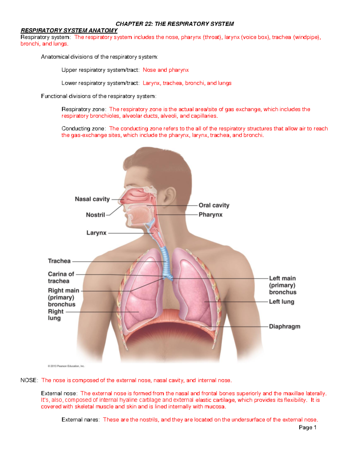
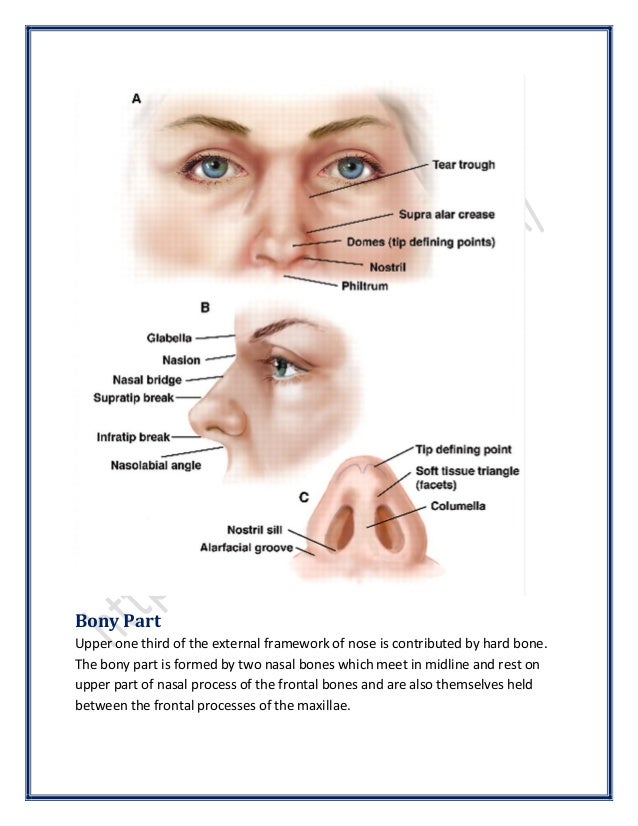
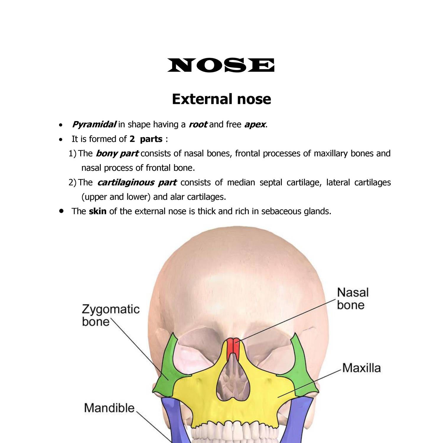
External nose anatomy. The external part of the nose includes the root between the eyes the dorsum that runs down the middle and the apex at the tip of the nose. The nasal root is located superiorly and is continuous with the forehead. The inferior portion of the nose is made up of hyaline cartilages.
The nose is a complex component of the facial anatomy that is comprised of numerous structures. The apex of the nose ends inferiorly in a rounded tip. The external nose is said to have a pyramidal shape.
It is partly formed by the nasal and maxillary bones which are situated superiorly. Anatomy of the nose. Lateral major alar minor alar and the cartilaginous septum.
Triangular shaped projection in the center of the face. The external skeleton extends the nasal cavities onto the front of the face see figure 1. Learn vocabulary terms and more with flashcards games and other study tools.
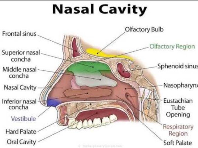
The nose is the external protuberance of an internal space the nasal cavity. It is subdivided into a left and right canal by a thin medial the nose has two cavities separated from one another by a wall of cartilage called the septum. External nose the nasal root is the top of the nose that attaches the nose to the forehead.
Osteocartilaginous framework upper one third is bony lower two third is cartilaginous 7. The cartilage also gives shape and support to the outer part of the nose. Here we will go from top to bottom and describe each of the components.
Made up mainly of cartilage and bone and covered by mucous membranes. Externally the nose is primarily a three sided pyramid. The noses exterior anatomy includes the nasal cavity paranasal sinuses nerves blood supply and lymphatics.
Composed of bones and cartilages the framework of the nose is covered with skin that is lined with mucous membrane. Start studying external nose anatomy. External nasal anatomy lets start with the external anatomy of the nose.
External nose the external nose has two elliptical orifices called the naris nostrils which are separated from each other by the nasal septum. 11 the nasal root is above the bridge and below the glabella forming an indentation known as the nasion at the frontonasal suture where the frontal bone meets the nasal bones. Two chambers divided by the septum.
The lateral margin the ala nasi is rounded and mobile.
 Does The Nose Contain Elastic Cartilage Quora
 38 Aesthetic Alteration Of The Nose Evaluation And Surgery
 Anatomic Considerations Semantic Scholar
 Nose Anatomy
 Rhinoplasty Michigan Manual Of Plastic Surgery Lippincott
 Nose Definition Anatomy Functions Diagram
 Anatomy Of External Nose By Av Sharma
 Dysmorphology Nose Flashcards Quizlet
 Surgical Treatment Of Nasal Obstruction In Rhinoplasty
 Regional Block Of The Nose Anatomy The Innervation Of The Nose
 Nasal Cavity
 Nasal Anatomy Dr Evan Ransom
 Anatomy Of External Nose By Av Sharma
 Anatomy Of The Nose
0514 Lateral View Of External Nose Anatomy Of Nasal Skeleton
 Anatomy Of Nose Steemit
 Nasal Cavities And Paranasal Sinuses Anatomy 1 With Luizzi
 Ch 22 Lecture Outline Bio 2063 Utsa Studocu
 Anatomy Of The Nose Surgicomed Com
 3 Lateral View Of The External Nose Showing The Cartilage
 Nostrils Definition Functions Anatomy Pictures
 Anatomy Of External Nose By Av Sharma
 Nose Structures External Nose Cartilage Philtrum Naris
 Head And Neck Anatomy Qa
 Anatomy Nose Doc Docdroid
 Nasal Cavity And Paranasal Sinuses Flashcards Quizlet
 0514 Lateral View Of External Nose Anatomy Of Nasal Skeleton
 Easy Notes On Nose Learn In Just 4 Minutes Earth S Lab
Ch 22 Lecture Outline Bio 2063 Utsa Studocu
Belum ada Komentar untuk "External Nose Anatomy"
Posting Komentar