Knee Anatomy Lateral
It is made of fibrocartilage. They are they soft tissues found at the end of muscles which link the muscle to bone.
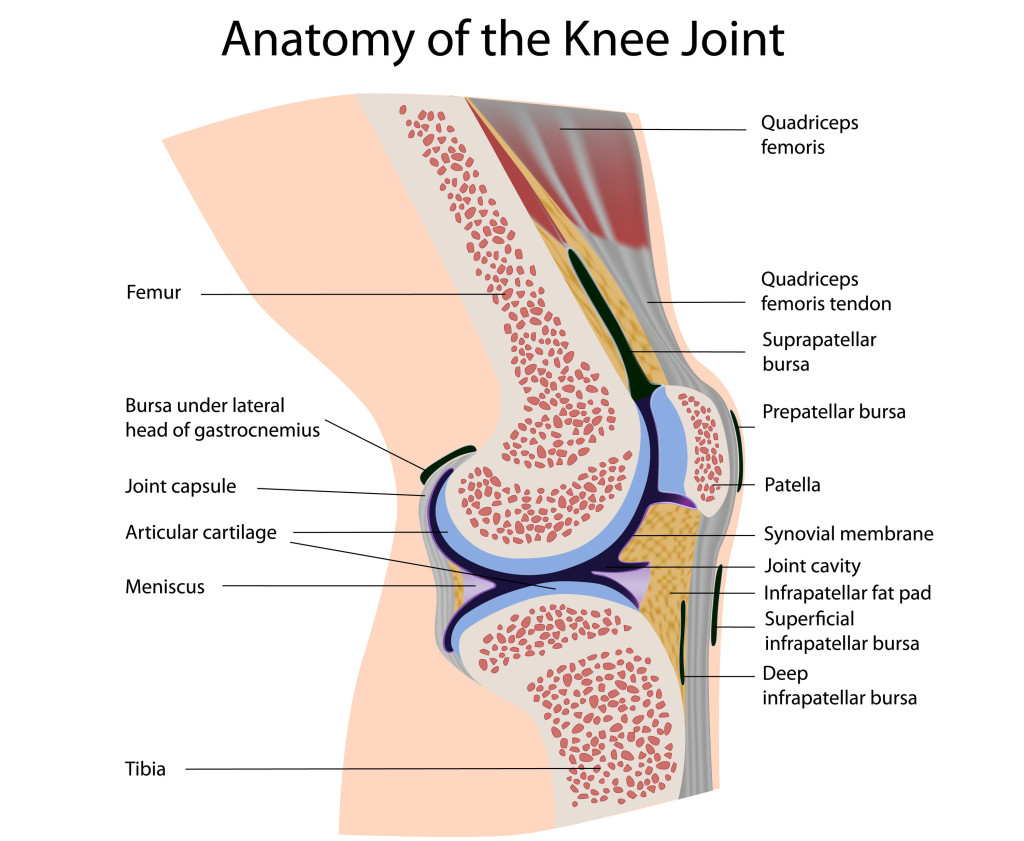
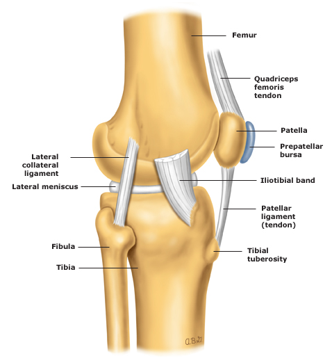
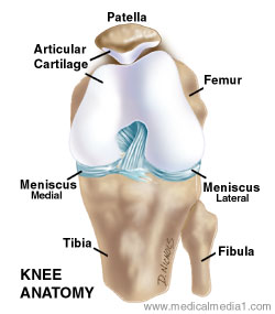
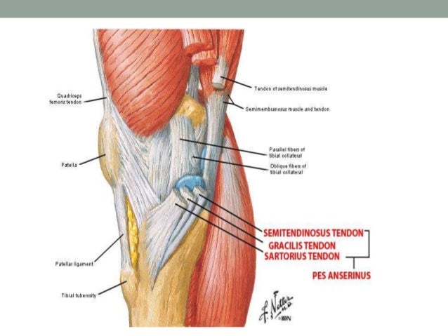
The knee cap actually sits inside the patellar tendon.
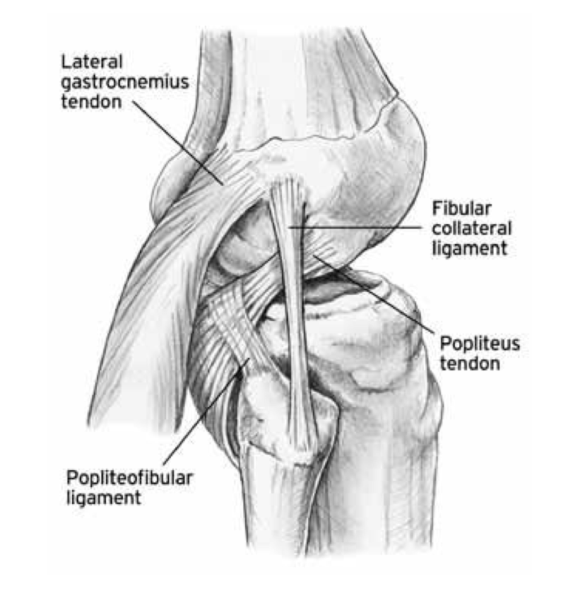
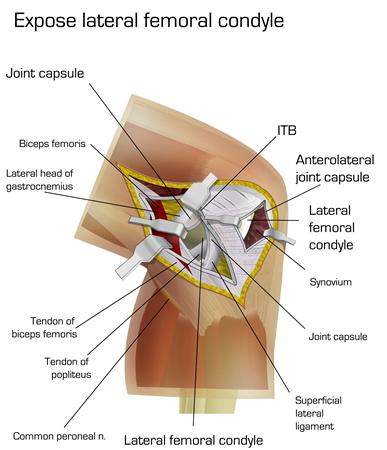
Knee anatomy lateral. Tendons at the knee. Tendons are often overlooked as part of knee joint anatomy. Lateral knee anatomy the lateral compartment of the knee contains several ligamentous and tendinous structures that are the first restraint against varus angulation and external internal rotation and anterior posterior translation of the leg bone.
A lack of familiarity leads to hesitancy when performing approaches in these areas of the knee. To act as shock absorbers by increasing surface area to further dissipate forces. It is the largest synovial joint in the body and allows flexion and extension of the leg as well as some rotation in the flexed position.
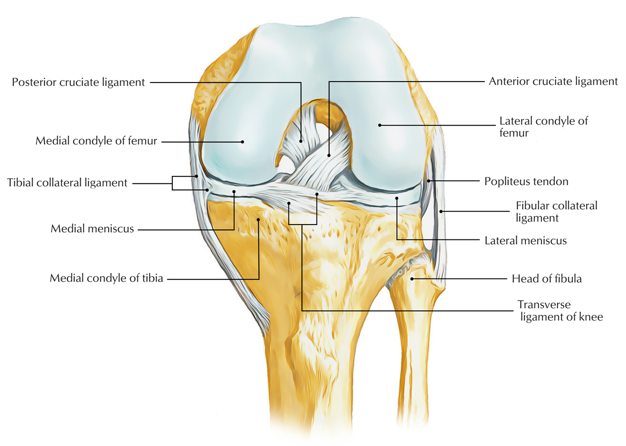
The bony shape of the posterolateral knee with the two convex opposing surfaces of the lateral femoral condyle and the lateral tibial plateau makes this portion of the knee inherently unstable compared to the medial aspect. It is attached to the tibia as well as to the joint capsule of the knee. It acts as a shock absorber in the knee and adds stability to the knee joint.
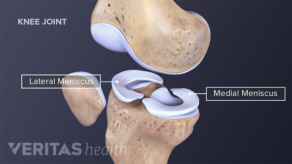
Knowledge of the bony topography will result in a greater number of anatomic ligament reconstructions. To deepen the articular surface of the tibia thus increasing stability of the joint. The medial meniscus is a crescent shaped structure that exists on the inside of the knee.
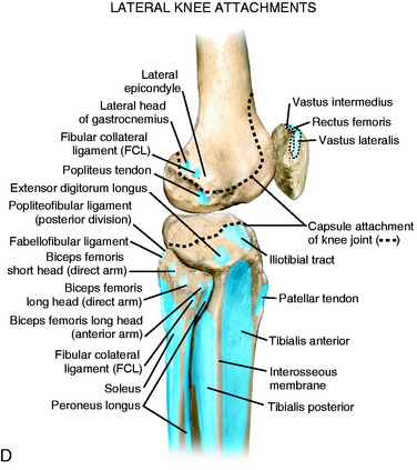
The main tendon found at the knee is the patellar tendon which links the quads muscles to the shin bone. Lateral knee bony anatomy the bony anatomy of the lateral knee is essential for understanding not only key relationships of soft tissue structures but also functions as a key determinant of the inability of many lateral knee injuries to heal over time. The posterior and lateral anatomy of the knee joint presents a challenge to even the most experienced knee surgeon.
The posterior and lateral anatomy of the knee joint presents a challenge to even the most experienced knee surgeon. The lateral meniscus sits on the lateral tibial plateau. Knowledge of the bony topography will result in a greater number of anatomic ligament reconstructions.
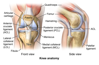
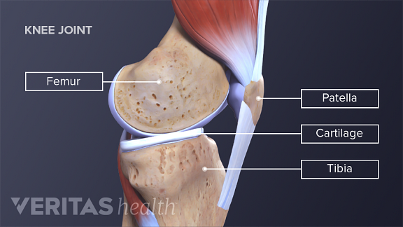
Two c shaped pieces of cartilage called the medial and lateral menisci act as shock absorbers between the femur and tibia. Soft tissue structures of the lateral knee attach to the distal femur. The knee joint is a modified hinge joint between the femur tibia and patella.
Numerous bursae or fluid filled sacs help the knee move smoothly. The medial and lateral menisci are fibrocartilage structures in the knee that serve two functions. Thus it has a much higher risk of not healing properly after injury than the medial aspect of the knee.
A lack of familiarity leads to hesitancy when performing approaches in these areas of the knee.
![]() Leg And Knee Anatomy Bones Muscles Soft Tissues Kenhub
Leg And Knee Anatomy Bones Muscles Soft Tissues Kenhub
 Knee Human Anatomy Function Parts Conditions Treatments
 The Knee Resource Posterolateral Corner Injury
 Lateral Posterior And Cruciate Knee Anatomy Clinical Gate
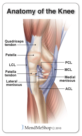
 Anatomy Of The Knee Central Coast Orthopedic Medical Group
 Redding Hospital Knee Anatomy
Common Knee Injuries Orthoinfo Aaos
 Lateral Knee Pain Pain On Outside Of Knee Knee Pain Explained


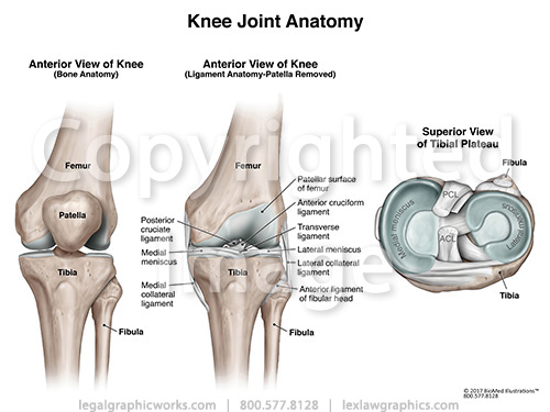 Anterior Tibial View Of Knee Joint Anatomy
 Lateral Medial And Posterior Knee Pain Brukner Khan S

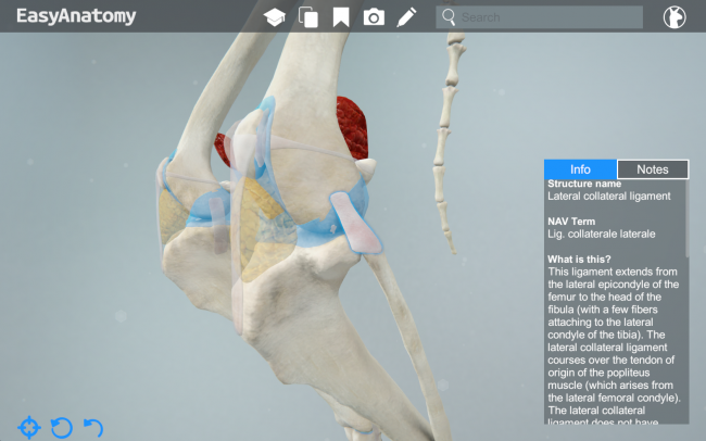
 Lateral Anatomy Knee Joint Uptodate
 Human Knee Joint Set 3d Illustration Stock Illustration
Easy Notes On Ligaments Of The Knee Joint Learn In Just 3
 Isolated Human Knee Anatomy Lateral View
 Lateral Outside Knee Pain Causes Treatment Your
 Knee Anatomy Dorsal View Stock Illustration
 Knee Wikipedia
 Knee Joint Anatomy Lateral View
 Anatomy Of The Knee Joint Paley Orthopedic Spine Institute
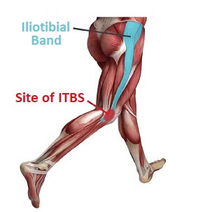
 Lateral Knee Injury Don T Get Sidelined By Iliotibial Band
 Knee And Related Knee Anatomy Images And Medical
/188058334-crop-56aae7425f9b58b7d0091480.jpg) What Is Causing Your Knee Pain
What Is Causing Your Knee Pain
 Ligaments Of The Knee Knee Sports Orthobullets
 Knee Anatomy
 Derived Copy Of Anatomy Of Selected Synovial Joints
 What S Causing My Knee Pain
 Lateral Approach To The Knee Approaches Orthobullets
 Anatomy And Examination Of The Knee
Belum ada Komentar untuk "Knee Anatomy Lateral"
Posting Komentar