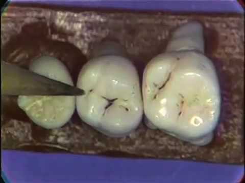
Anatomy Of Molar Teeth
Molars 8 total. Gross anatomy there are twenty deciduous primary teeth in young children with ten per jaw and five in each quadrant which consist of distal to mesial.
 Stock Illustration
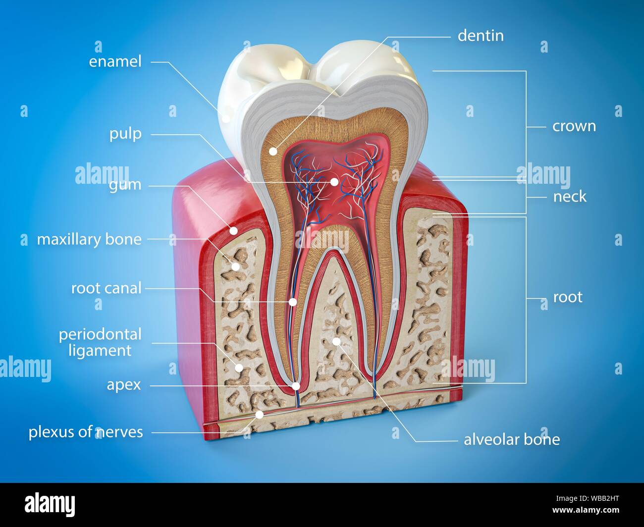
Knowing the tooth anatomy will help you understand how dental problems occur and how the treatment is performed.
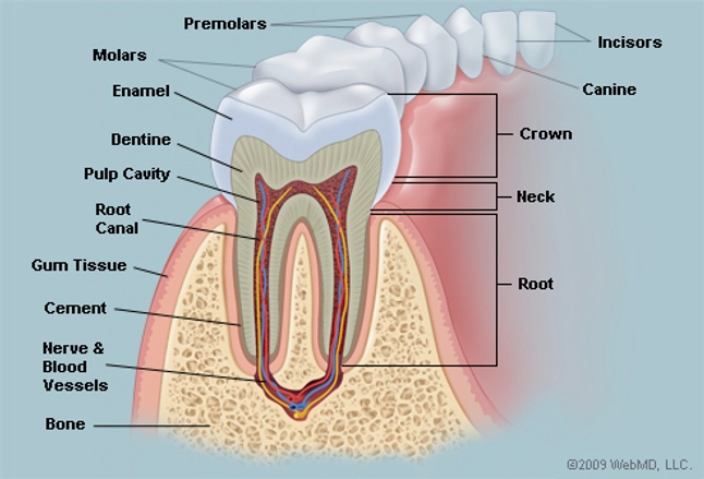
Anatomy of molar teeth. The function of teeth as they contact one another falls elsewhere under dental occlusion tooth formation begins before birth. The root canal is a passageway that contains pulp. The deciduous primary teeth start erupting at six months lower central incisor and are completely erupted by around 3 years of age.
Anatomy of the tooth the tooth is one of the most individual and complex anatomical as well as histological structures in the body. However they have the same tooth anatomy. Without proper brushing and flossing plaque and tartar can build up at the gumline leading to gingivitis and gum disease.
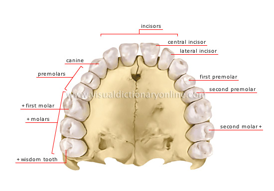
Wisdom teeth or third molars 4 total. What are the different parts of a tooth. Also every tooth made of several layers.
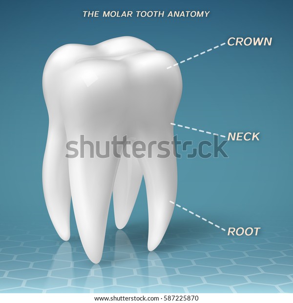
Teeth are different in shape. The tissue composition of a tooth is only found within the oral cavity and is limited to the dental structures. Every tooth consists of three parts.
For example front teeth are sharp and chisel shaped for cutting while molars have flat surfaces for grinding. The crown neck and root. The shape of the crown determines the tooths function.
Also called cement this bone like material covers the tooths root. Crown the top part of the tooth and the only part you can normally see. Flat teeth in the rear of the mouth best at grinding food.
An tooth is an unerupted or partially erupted tooth that is positioned against another tooth bone or even soft tissue in such a way that only partial eruption is likely if at all anodontia permanent maxillary third molars along with the mandibular third molars commonly exhibit partial hypodontia and thus are congenitally. It makes up approximately two thirds of the tooth. The root is the part of the tooth that extends into the bone and holds the tooth in place.
Peaks and valleys on the flat apical surface of premolars and molars are used for chewing and grinding food into tiny pieces. Its made up of several parts. The development appearance and classification of teeth fall within its purview.
These teeth erupt at around age 18 but are often surgically removed. Dental anatomy is a field of anatomy dedicated to the study of human tooth structures. Premolars bicuspids and molars are large flat surfaced teeth found in the back of the mouth.
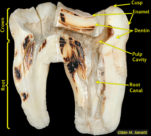
The enamel dentin cementum and pulp. Gumline where the tooth and the gums meet.
 The Teeth Human Anatomy Diagram Names Number And
 Molar Tooth Britannica
 Amazon Com Dental Teeth Anatomy Structure Molar Incisor 2
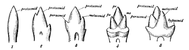
 Bio370 Mammal Teeth
 Molar Anatomy Crown Neck Root Tooth Stock Vector Royalty
 Root Canal Morphology And Variations In Mandibular Second
 Dental Anatomy Maxillary Molars
 Molar Tooth Cross Section Stock Photos Molar Tooth Cross


The Mouth Pharynx And Esophagus Anatomy And Physiology

 Dental Tooth Oclusal Stock Illustration Illustration Of

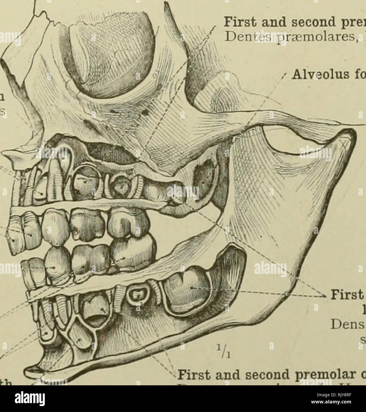 An Atlas Of Human Anatomy For Students And Physicians

 Molar Tooth Wikipedia
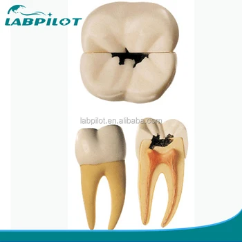 Molar Tooth Decay Anatomical Model Dental Caries Model Buy Dental Caries Tooth Decay Molar Product On Alibaba Com
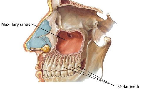 Sinus Lifts And Dental Implants Dr Nima Massoomi Dmd Med Md
 External And Internal Root Canal Anatomy Of The First And
 Pdf Evaluation Of Complex Mesiobuccal Root Anatomy In
 Molar Teeth Diagram Types Of Electrical Wiring Diagrams
 Dental Anatomy Permanent Molars

Anatomy And Development Of The Mouth And Teeth
Belum ada Komentar untuk "Anatomy Of Molar Teeth"
Posting Komentar