Basal Ganglia Anatomy
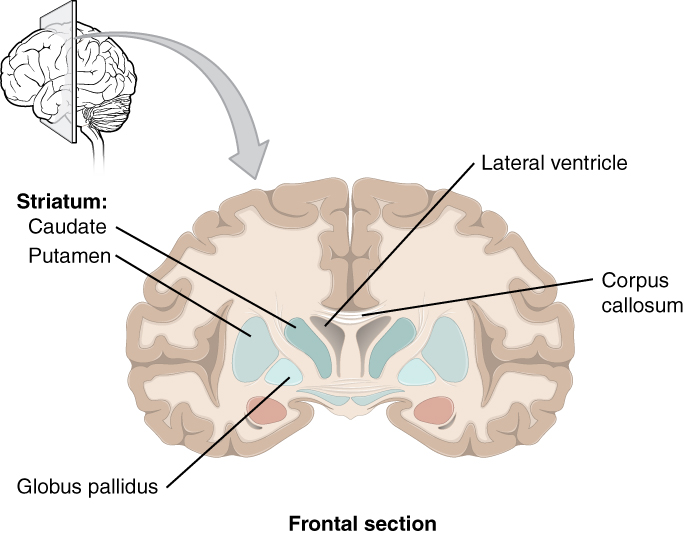
The term basal ganglia usually includes the caudate putamen globus pallidus and amygdala. The basal ganglia are a group of grey matter nuclei in the deep aspects of the brain that is interconnected with the cerebral cortex thalami and brainstem.
 The Basal Ganglia Purposegames
Anatomically speaking this brain structure has four parts or distinct nuclei.
Basal ganglia anatomy. The basal ganglia are involved primarily in processing movement related information. Amygdaloid nuclear complex or amygdala. In a strict anatomical sense it contains three paired nuclei that together comprise the.
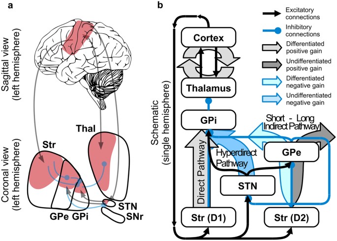
Anatomically the basal ganglia consist of parallel complementary pathways. The basal ganglia is composed of the following grey nuclei. The basal ganglia nuclei of the basal ganglia.
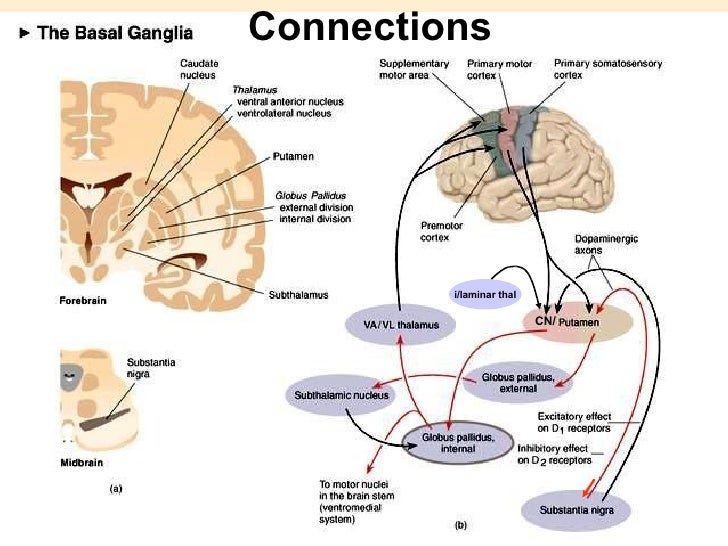
Basal ganglia snc and cm pf nuclear complex connections pallidum striatum thalamus cm pf pallidum striatum snc 19. We use the term more loosely to refer to a group of nuclei that are anatomically interconnected and have important motor functions. The majority of basal ganglia nuclei have projection neurons.
Two of them the striatum and the pallidum are relatively large. Basal ganglia oculomotor loop connections frontal eye field area 8 primary motor area m i thalamus vlm vamc md striatum caudate nucleus snr substantia nigra pars reticulata pyramidal tract lmn tectum 18. The basal ganglia consist of the corpus striatum a major group of basal ganglia nuclei and related nuclei.
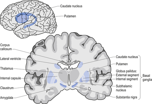
Substantia nigra within the midbrain. The anatomy of the basal ganglia is complex since it is spread throughout. In terms of anatomy the basal ganglia are divided into four distinct structures depending on how superior or rostral they are in other words depending on how close to the top of the head they are.
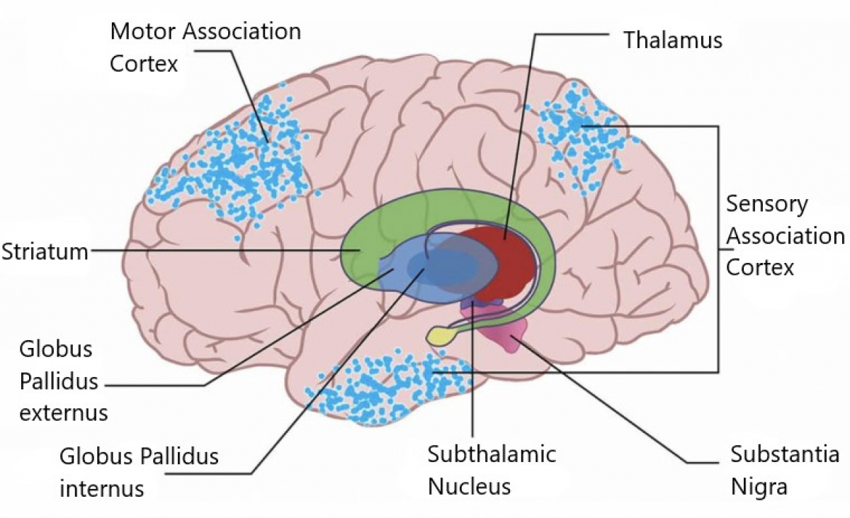
Basal ganglia anatomy and connections. The basal ganglia are a group of neurons also called nuclei located deep within the cerebral hemispheres of the brain. In simple terms the basal ganglia provide a feedback mechanism to the cerebral cortex.
Basal ganglia anatomy as previously mentioned basal ganglia are fundamental brain structures that assemble different gray matter nuclei stored in the deepest regions of the brain. In order to execute purposeful movements a small number. These are the caudate putamen globus pallidus substantia nigra and subthalamic nuclei.
The other two the substantia nigra and the subthalamic nucleus are smaller.
 Anatomy Of Basal Ganglia
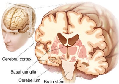
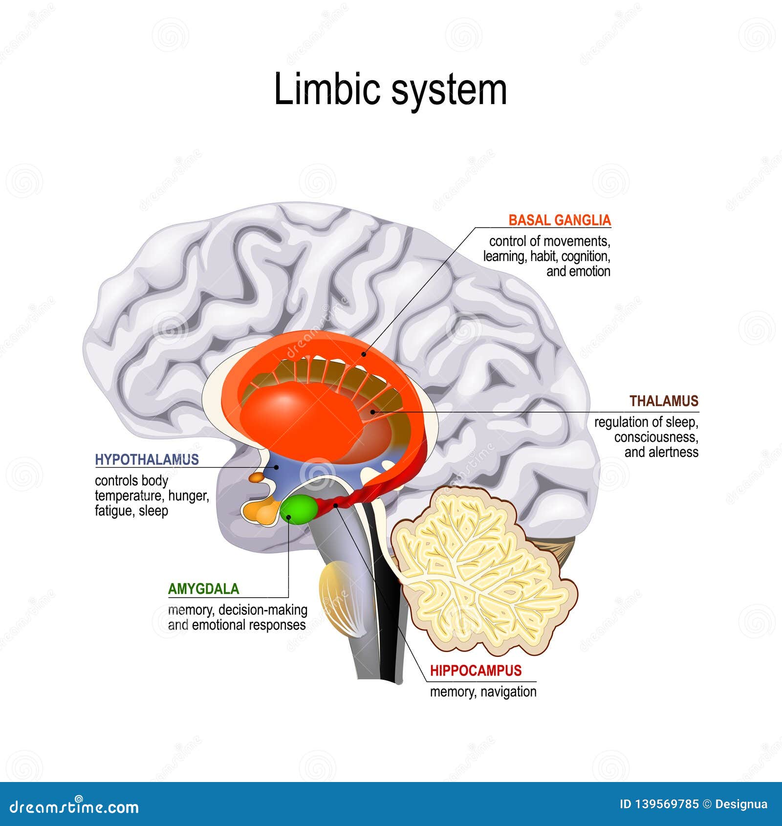 Limbic System Cross Section Of The Human Brain Stock Vector
 Basal Ganglia Fill In The Blanks Sketchy Medicine
 Basal Nuclei Human Anatomy
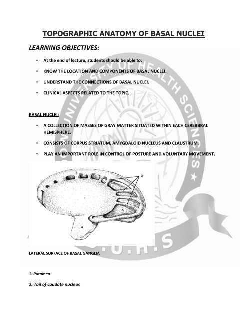 Topographic Anatomy Of Basal Nuclei
 An Introduction To The Basal Ganglia Fewer Lacunae
 Telencephalon Language Centers Limbic System Basal Ganglia
 Basal Ganglia And Thalamus Postgraduate Training
 Basal Ganglion An Overview Sciencedirect Topics
 Basal Ganglia Basal Nuclei Function Anatomy Clinical
 Basal Nuclei Human Anatomy Organs
 The Basal Ganglia Clinical Gate
 Changing Pattern In The Basal Ganglia Motor Switching Under
 Ppt Basal Ganglia Powerpoint Presentation Free Download
 Basal Ganglia A Definition Anatomy And Function
 Neuroanatomy Lectures Basal Ganglia Part 1 Anatomy
 Myneurologytips Basal Ganglia Anatomy
 Control Circuits Integrate And Control The Activities Of The
![]() Basal Ganglia Anatomy Of Direct And Indirect Pathways Kenhub
Basal Ganglia Anatomy Of Direct And Indirect Pathways Kenhub
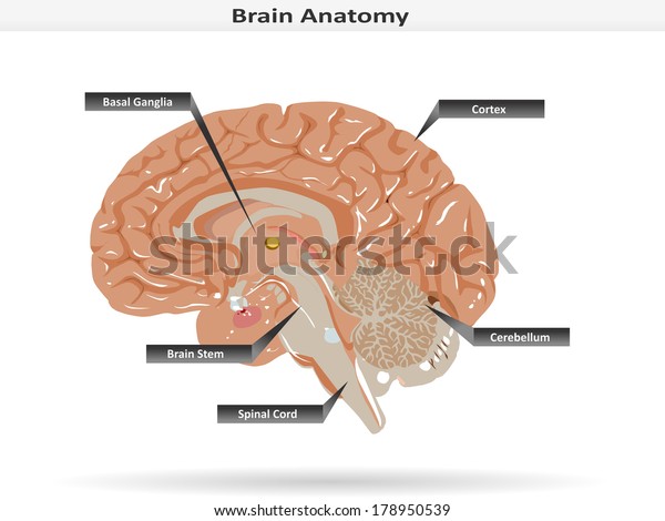 Brain Anatomy Basal Ganglia Cortex Brain Stock Vector
 Drawing Of The Brain Showing The Basal Ganglia Abd Thalamic Nuclei
 Basal Ganglia Pharmacology Sammy Case Matt Vreugde Ppt
![]() Basal Ganglia Anatomy Of Direct And Indirect Pathways Kenhub
Basal Ganglia Anatomy Of Direct And Indirect Pathways Kenhub
 Basal Ganglia Anatomy Function Stroke And Disorders
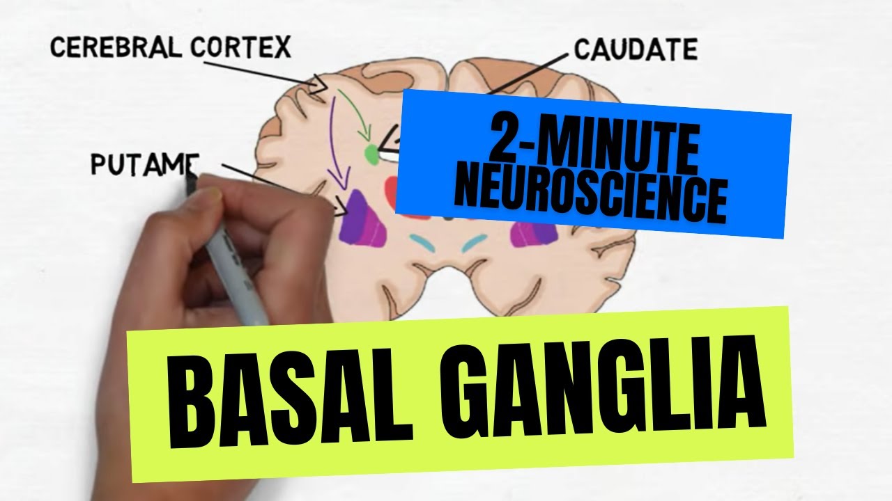
2 Minute Neuroscience Basal Ganglia
 Thalamus And Basal Ganglia Radiology Key

Belum ada Komentar untuk "Basal Ganglia Anatomy"
Posting Komentar