Anatomy Of Orbit
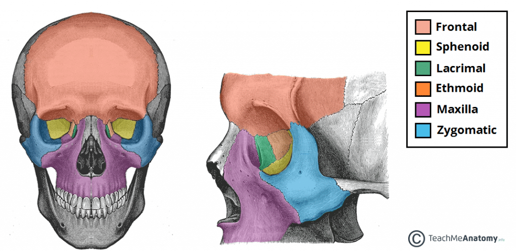
Anatomy of the eye and orbit. Development orbit develops around the eyeball orbital walls derived from cranial neural crest cells which expand to form frontonasal process maxillary process lateral nasal process maxillary process medial inferior and lateral orbital walls capsule of forebrain forms orbital roof.
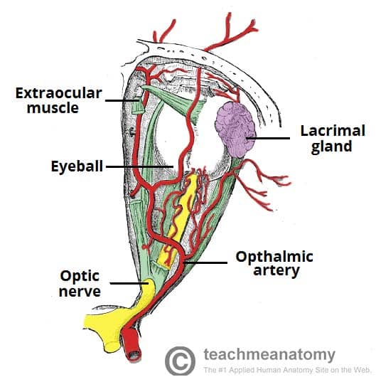
 Anatomy Of The Orbit Vessels And Nerves
101 us fl oz.
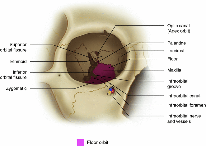
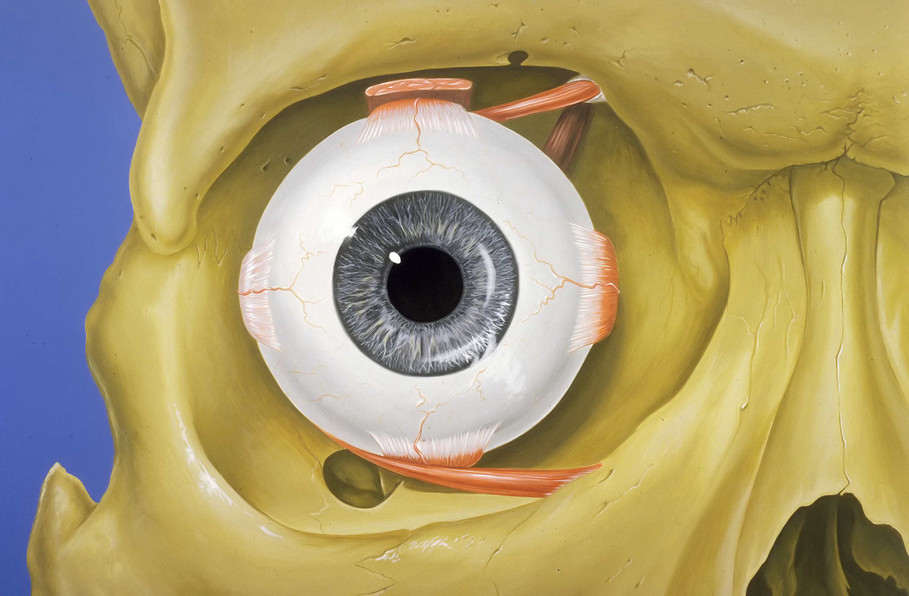
Anatomy of orbit. The shape of the orbit resembles a four sided pyramid to begin with but as one goes posterior it becomes three sided towards the apex. The orbit which protects supports and maximizes the function of the eye. The clinical essentials achieves the impressive task of presenting ophthalmology residents optometry residents and optometry students with the clinical essentials of ocular anatomy as a foundation for patient care.
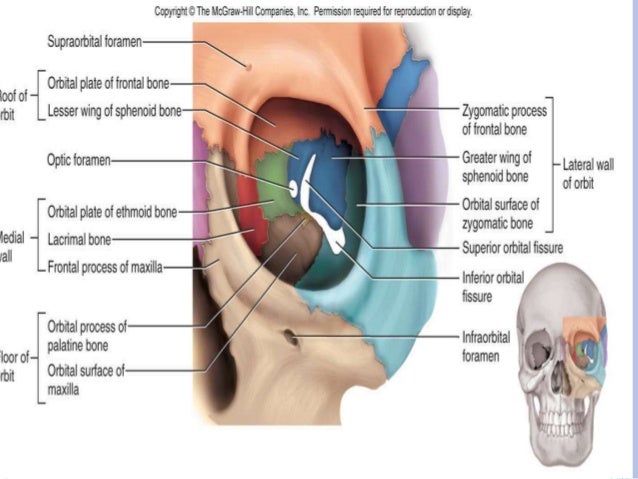
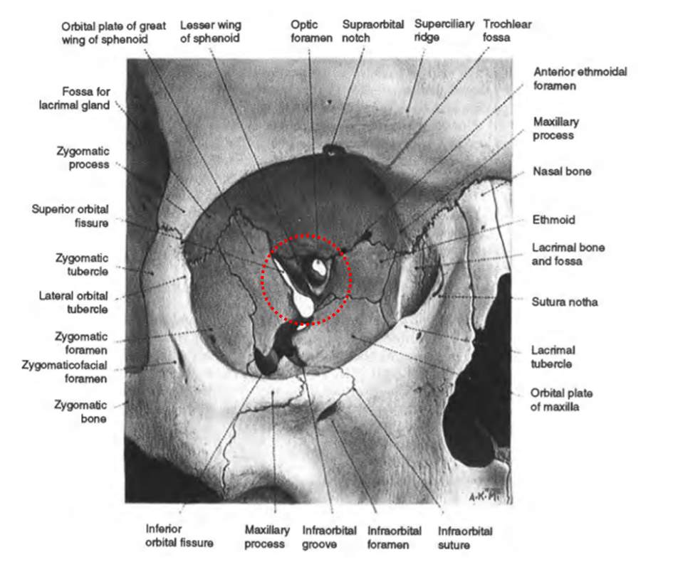
It emphasizes the aspects of eye and orbit anatomy that are most relevant to clinicians in training providing the practical real world foundation necessary for practice. Borders of orbit roof floor base apex medial and lateral walls of orbit superior orbital fissure inferior orbital fissure superior orbital foramen inferior orbital foramen optic. Orbit supports the eye and ensures that this organ functions in an optimal manner.
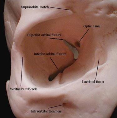
Orbit anatomy in anatomy the orbit is the cavity or socket of the skull in which the eye and its appendages are situated. The lacrimal system produces distributes and drains tears. In the adult human the volume of the orbit is 30 millilitres 106 imp fl oz.
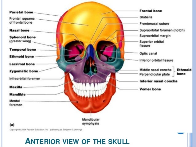
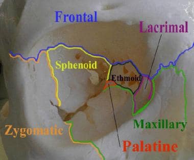
Anatomy of the orbit the skull is composed of two segments the cranium and the face. The cranium is the major portion and it consists of three unpaired bones the sphenoid occipital and ethmoid bones and three paired bones the frontal parietal and temporal bones. The volume of the orbital cavity in an adult is roughly about 30cc.
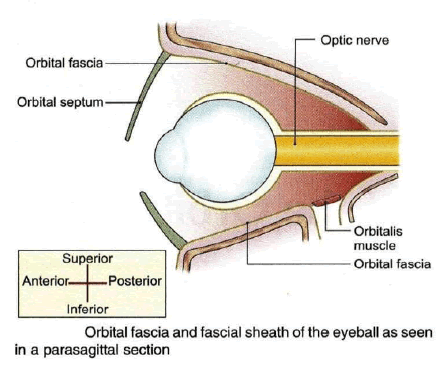
It also protects this vital structure. When orbital cellulitis occurs its most likely source is direct extension from the ethmoid sinuses because the thin bone of the medial wall is easily penetrated by expanding masses from the sinus. The contents of the orbit are separated and supported by multiple.
The floor of the orbit is thicker and offers more resistance to maxillary sinus abnormality. Orbit can refer to the bony socket or it can also be used to imply the contents.
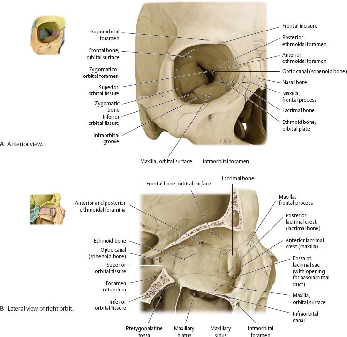
![]() Bones Of The Orbit Anatomy Foramina Walls And Diagram
Bones Of The Orbit Anatomy Foramina Walls And Diagram
 The Bony Orbit Borders Contents Fractures Teachmeanatomy
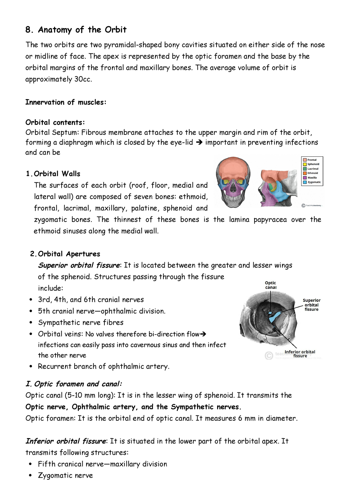 8 Anatomy Of Orbit Grade 5 Ophthalmology Studocu
 Orbital Bone Anatomy Eye Anatomy Facial Anatomy
 File 1411 Eye In The Orbit Jpg Wikimedia Commons
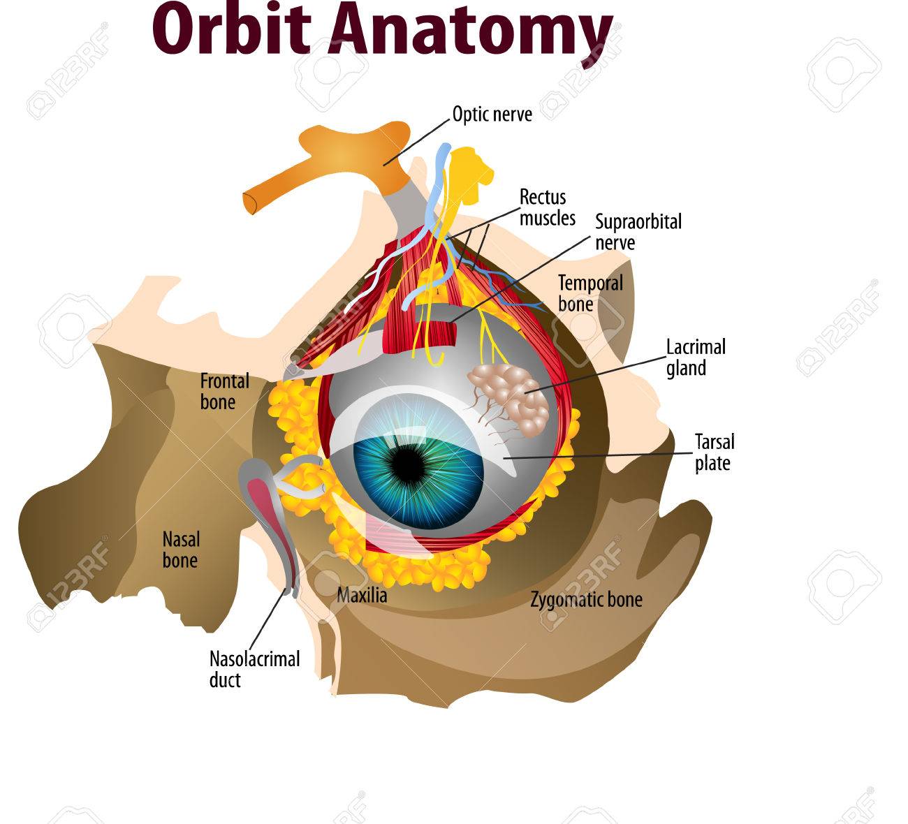 Vector Illustration Of Orbit Anatomy
 Normal Orbital Anatomy Axial Computed Tomographic Ct
 Eye Orbit Eye Anatomy Orbit Anatomy Anatomy
 Anatomy Of The Orbit Springerlink
 Anatomy Of The Orbit
 Anatomy Of The Eye Orbit Alila Medical Images
 Anatomy Of The Orbit
 Orbit Anatomy Osteology Lacrimal System Connective Tissue
 Ocular Orbit
 Anatomy Of The Posterior Orbit And Orbital Apex
 Applied Anatomy Of Orbit Download Table
 Orbital Region
 Orbital Anatomy Tutorial
![]() Bones Of The Orbit Anatomy Foramina Walls And Diagram
Bones Of The Orbit Anatomy Foramina Walls And Diagram
 Periorbita An Overview Sciencedirect Topics
 Orbit Anatomy Wikipedia
 The Bony Orbit Borders Contents Fractures Teachmeanatomy
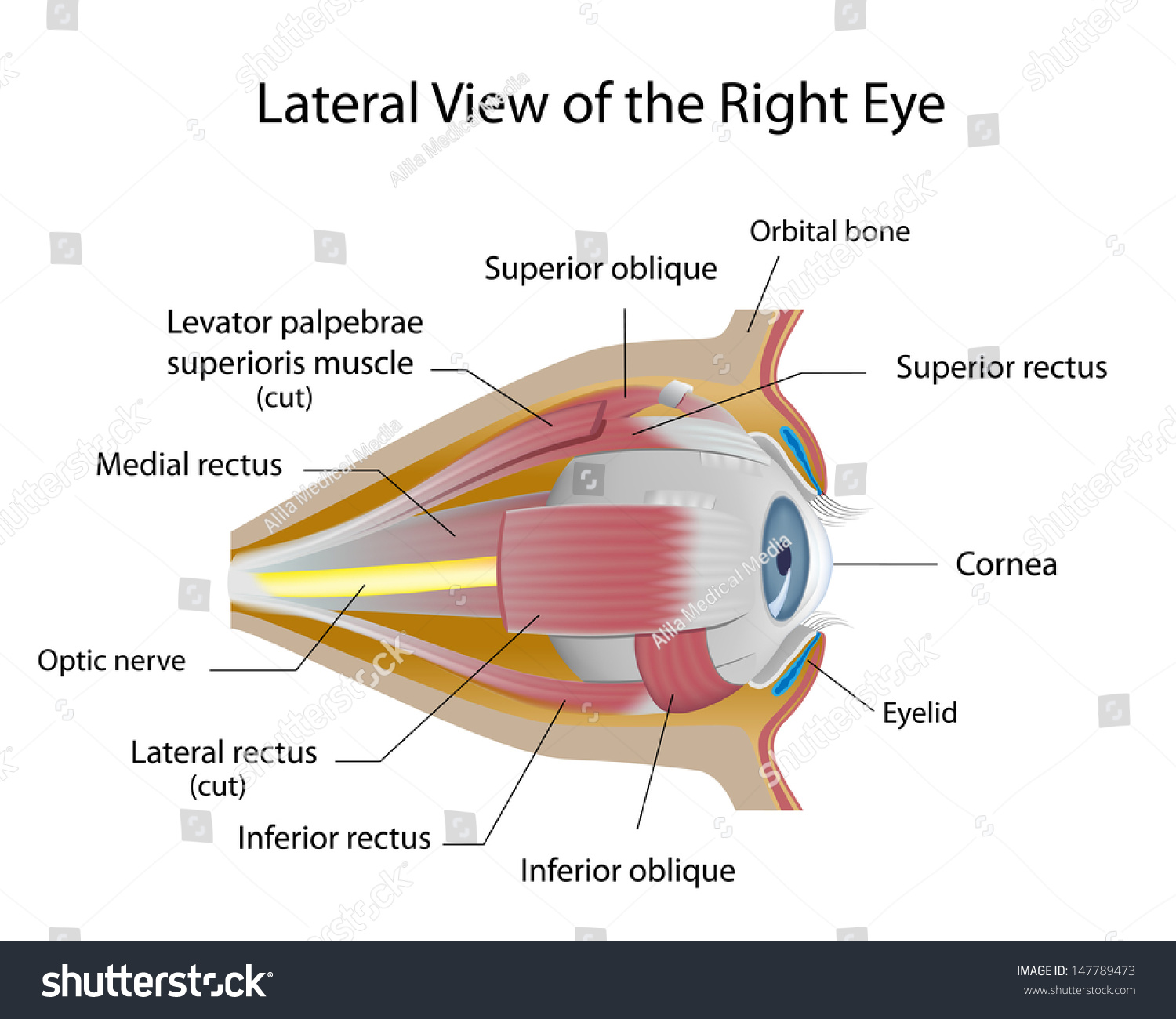 Human Eye Orbit Anatomy Stock Illustration 147789473
 Anatomy Of The Eye And Orbit Ento Key


 Orbit Anatomy Purposegames
 Instant Anatomy Diagram
 Orbital Tumor Eye Socket Cancer Anatomy
 Orbit Anatomy Osteology Lacrimal System Connective Tissue
Orbit Eye Atlas Of Anatomy
Belum ada Komentar untuk "Anatomy Of Orbit"
Posting Komentar