C1 And C2 Anatomy
The atlas and axis are specialized to allow a. The second cervical vertebrae c2 is known as the axis.
 Cervical Vertebra An Overview Sciencedirect Topics
Together they form the atlantoaxial joint which is a pivot joint.
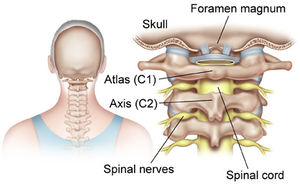
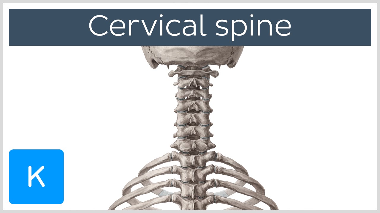
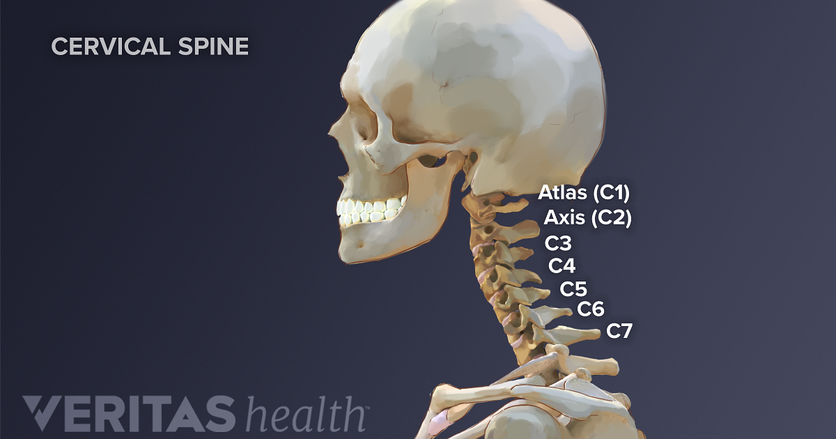
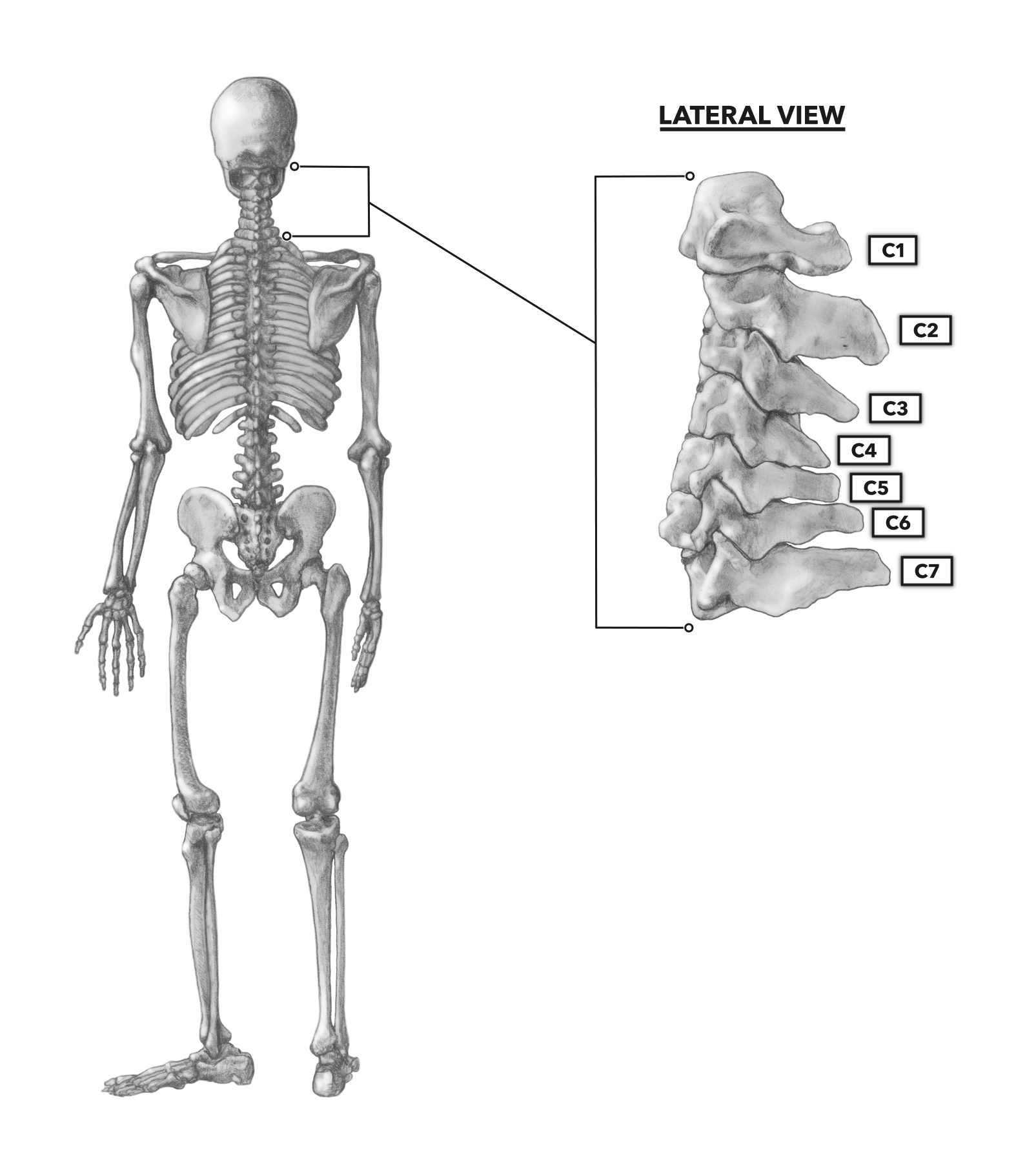
C1 and c2 anatomy. The purpose of the cervical spine is to contain and protect the spinal cord support the skull and enable diverse head movement. The first cervical vertebrae c1 is known as the atlas. Surgery of the c1 c2 vertebral andor spinal level is usually considered in one or more of the following cases.
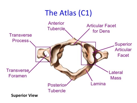
The c1 vertebra connects the skull to the cervical spine. The atlanto axial articulation is a complex of three synovial joints which join the atlas c1 to the axis c2. C1 serves as a ring or washer that the skull rests upon and articulates in a pivot joint with the.
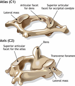
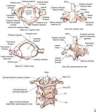
The c1 vertebra is formed like a ring that sits on top of c2. The axis is the second cervical vertebra commonly called c2. The c1 and c2 vertebrae are the first two vertebrae at the top of the cervical spine.
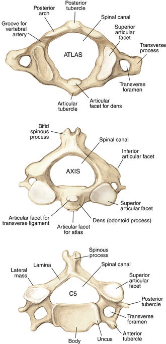
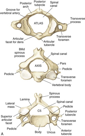
The individual cervical vertebrae are abbreviated c1 c2 c3 c4 c5 c6 and c7. C1 and c2 form a unique set of articulations that provide a great degree of mobility for the skull. Gross anatomy articulations paired lateral atlanto axial joints.
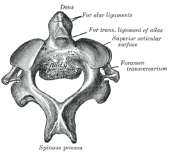
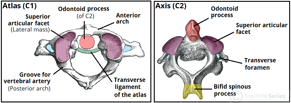
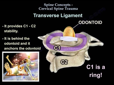
The c2 vertebra has a bony knob that fits into the front portion of the ring of the c1 vertebra. This bony knob is called the odontoid process or dens. The cervical nerves are also abbreviated.
In anatomy the atlas c1 is the most superior first cervical vertebra of the spine. The atlas is the topmost vertebra and with the axis forms the joint connecting the skull and spine. It is named for the atlas of greek mythology because it supports the globe of the head which is the skull.
Classified as planar type joint between the lateral masses of c1. Please support us patreon https. Its most prominent feature is the odontoid process or dens which is embryologically the body of the atlas c1 12.
These two vertebrae have different anatomy than the rest of the spine. They are c1 through c8. It is an atypical cervical vertebra with unique features and important relations that make it easily recognisable.
More of the heads rotational range of motion comes from c1 c2 than any other cervical joint. Join us in this video where we show the anatomy of atlas and axis c1 c2 through the use of models. The c1 sits atop and rotates around c2 below.
In this article we shall look at the anatomy of the cervical vertebrae their characteristic features articulations and clinical relevance. When the transverse ligamentligament that holds the c1 and c2 vertebrae together is partially or completely torn.
 The Spine Anatomy And Physiology
Acute Atlantoaxial Rotary Subluxation Aars Pediatric
 Rheumatoid Arthritis Of The Cervical Spine Redlands Loma
 Vertebrobasilar Insufficiency Hunter Bow Syndrome
 Spine Musculoskeletal Key
 Axis C2 Radiology Reference Article Radiopaedia Org
Axis Anatomy Wikipedia
 Spine Clinical Gate

 The Cervical Spine Features Joints Ligaments
 C1 And C2 Atlantoaxial Instability
 Anatomical Representation Of A Human Neck Illustrating Its
 Chapter 25 Overview Of The Neck The Big Picture Gross
 Cervical Vertebrae Physiopedia
 The C1 C2 Vertebrae And Spinal Segment
 Cervical Spine Sprain Strain Injuries Background
 Amazon Com Axis Scientific Occipital Bone C1 And C2
 C1 Vertebra Atlas And Accompanying Structures The Art Of
 Upper Cervical Spine Reduction Fixation Posterior C1
 Atlas Anatomy Cervical Vertebrae Anatomy Neck Anatomy
 Crossfit The Cervical Vertebrae
 Basic Spinal Anatomy Welcome Back Clinic Mri And Pain
Belum ada Komentar untuk "C1 And C2 Anatomy"
Posting Komentar