Anatomy Of An Ankle Sprain
After the examination your doctor will determine the. Grades of ankle sprains.

The name describes exactly where it is.
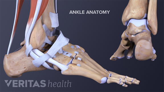
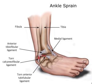
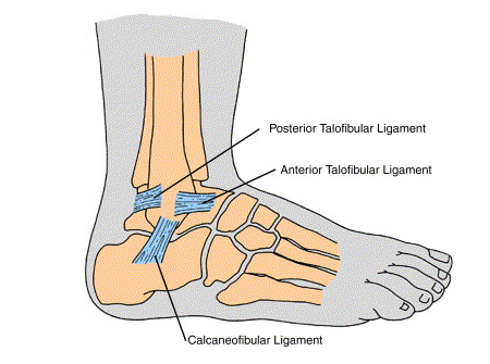
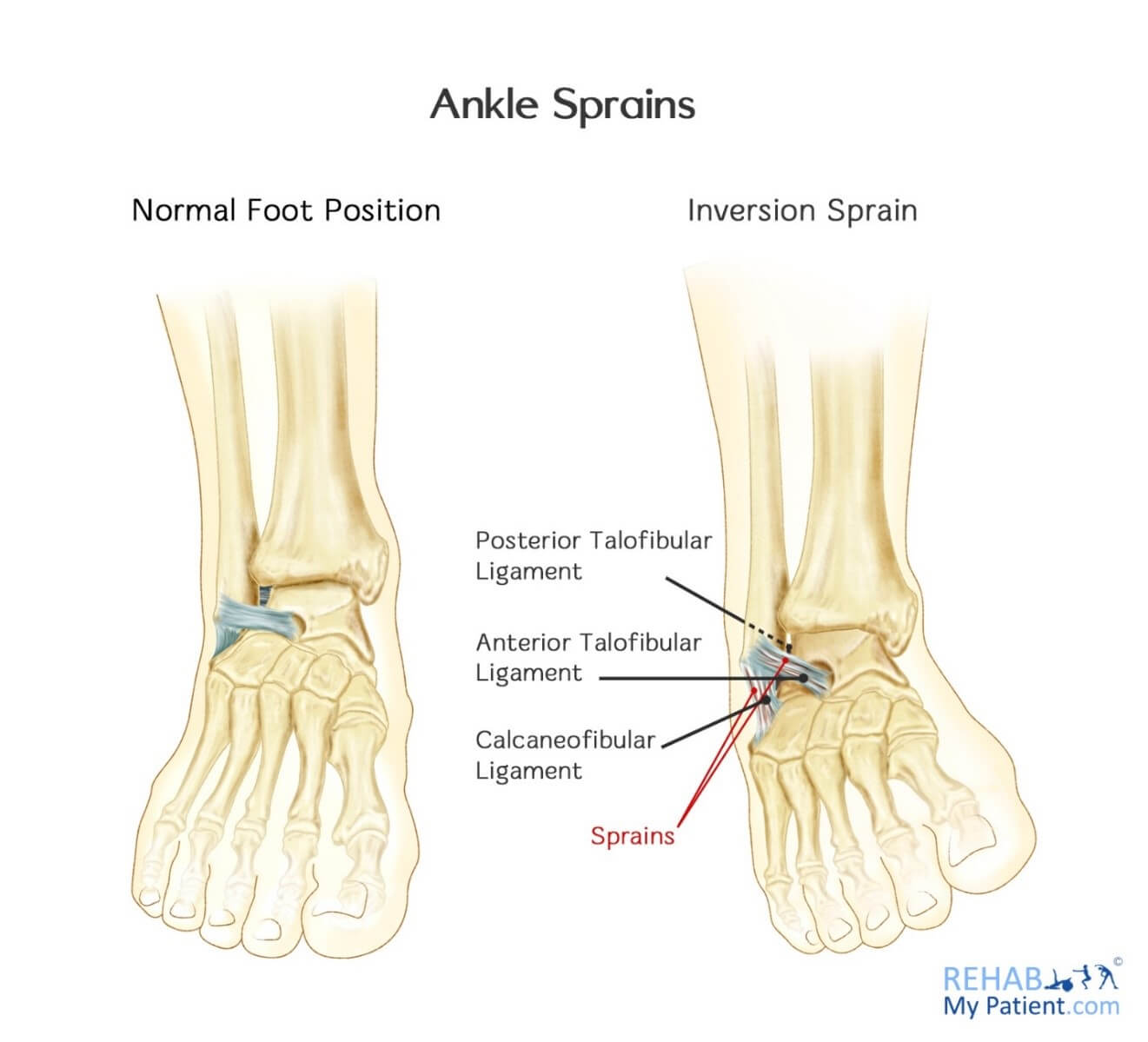
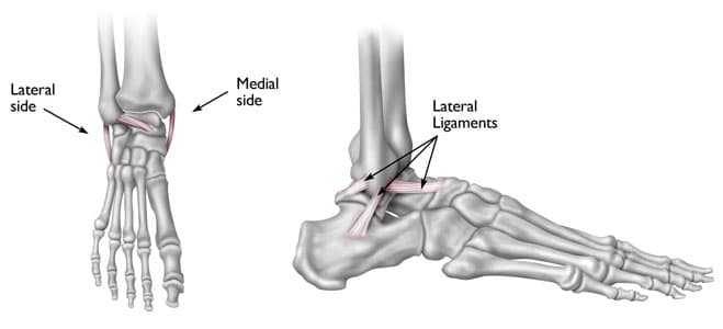
Anatomy of an ankle sprain. Normally dorsiflexion causes the interosseous ligament to become taut. In a typical lateral ankle sprain the most common ligament that is damaged is the anterior talofibular ligament. Your doctor will diagnose your ankle sprain by performing a careful examination.
Anatomy of an ankle sprain description. Widening of the ankle mortise that causes syndesmosis injury can also be the result of excessive or severe dorsiflexion. Over the counter as well as prescribed pain relief medications are fast.
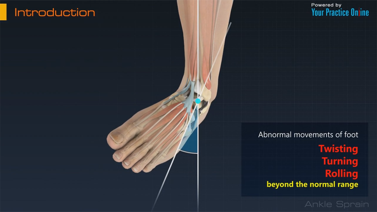
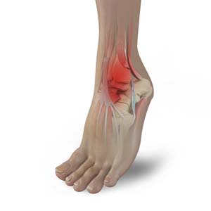
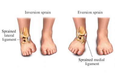
Damage to one of the ligaments in the ankle usually from an accidental twist. In a more severe sprain the calcaneofibular ligament may also be injured. Talar bone trauma obstructed by swelling.
An individual with an ankle sprain can almost always walk on the foot albeit carefully and with pain. These are the signs and symptoms of inflammation from the ankle sprain. The history of an ankle sprain is usually that of an inversion type twist of the foot followed by pain and swelling.
An ankle sprain occurs when the ligaments found in this joint are either partially or completely due to an accidental twist of the foot. It connects the talus bone of the ankle to the fibula in the lower leg. It is a synovial hinge joint.
This may occur in several sport activities or from a bad landing after a jump 4. A break in any of the three bones in the ankle. Tissue injury and inflammation occur when an ankle is sprained.
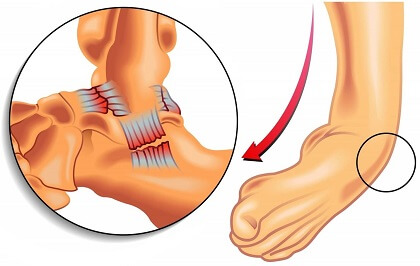
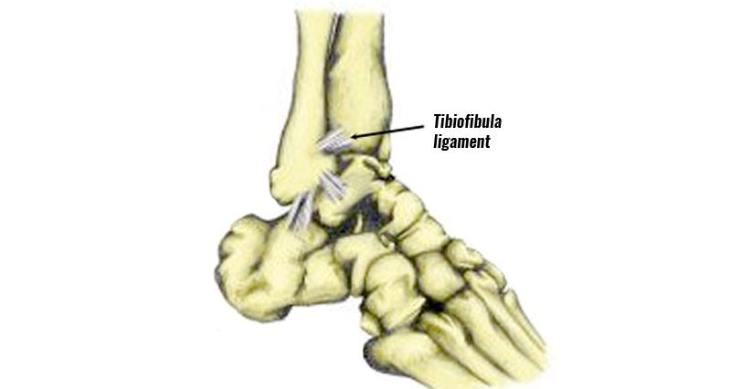
2 however since the anterior aspect of the dome of the talus is wider than the posterior aspect. Ankle anatomy of 3 common sprains. The ligament joining the two bones of the lower leg tibia and fibula.
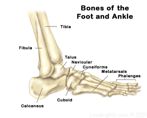
Ankle anatomy of sprains and breaks. The ankle joint and the subtalar joint. The ankle is made up of two joints.
Ankle conditions sprained ankle. Blood vessels become leaky and allow fluid to ooze into the soft tissue surrounding the joint. This is also called the ankle joint proper or the talocrural joint.
As illustrated here the three ligaments most commonly involved. Ligaments are strong fibrous tissues that connect bones to other bones. White blood cells responsible for inflammation migrate to the area and blood flow increases.
The ankle joint includes two bones the tibia and the fibula that form a joint that allows the foot to bend up and down.
 Twisted Ankle Symptoms Diagnosis Treatment Foot Pain
 Anatomy 101 Ankle Syndesmosis Distal Tibiofibular Joint

 All About Ankle Sprains And Strains
 Aspetar Sports Medicine Journal Ankle Sprain Diagnosis
 Ankle Sprain Rural Physio At Your Doorstep Physio Direct
 Anatomy 101 Ankle Syndesmosis Distal Tibiofibular Joint
 Ankle Sprains The Foot Practice Recommended Podiatrist
 Patient Education Article Familycare Of Kent
 Ankle Sprain Footcaremd
 Anatomy Of An Ankle Sprain
 Foot Ankle Trauma Miami Fl Sprains Strains Ankle
 Ankle Sprains Management
 Ankle Sprain Pinnacle Orthopaedics

 Ankle Sprain Foot Ankle Orthobullets
 High Ankle Sprain Syndesmotic Injury Footcaremd
 High Ankle Sprain Anatomy
 Ankle Wikipedia
 Anatomy Of An Ankle Sprain Bouldercentre For Orthopedics
 Ankle Sprain Physiopedia

 High Ankle Sprain Symptoms Causes Treatment And
 Why Ankle Pain Treatments Chronic Ankle Pain Ankle Joint
 Sprained Ankle Causes Symptoms Diagnosis Treatment

 Ankle Sprains
 Sprained Ankle Information And Treatment Suggestions For
 Inversion Sprain Of The Ankle Rehab My Patient
Ankle Sprain
 Anatomy Of An Ankle Sprain Bouldercentre For Orthopedics
 Ankle Sprains How To Take Care Of An Ankle Sprain
Belum ada Komentar untuk "Anatomy Of An Ankle Sprain"
Posting Komentar