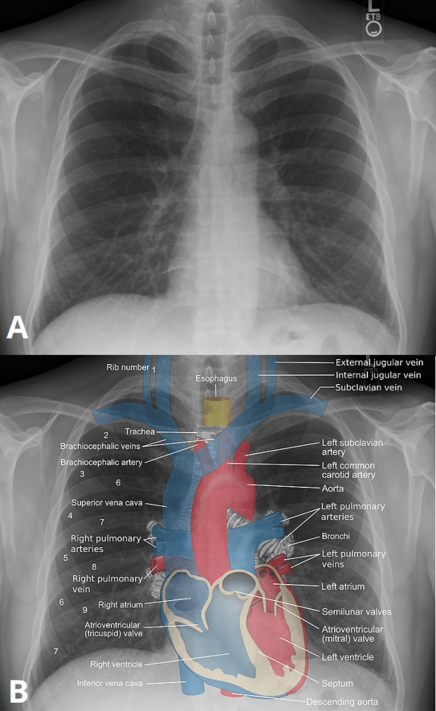
Shoulder X Ray Anatomy
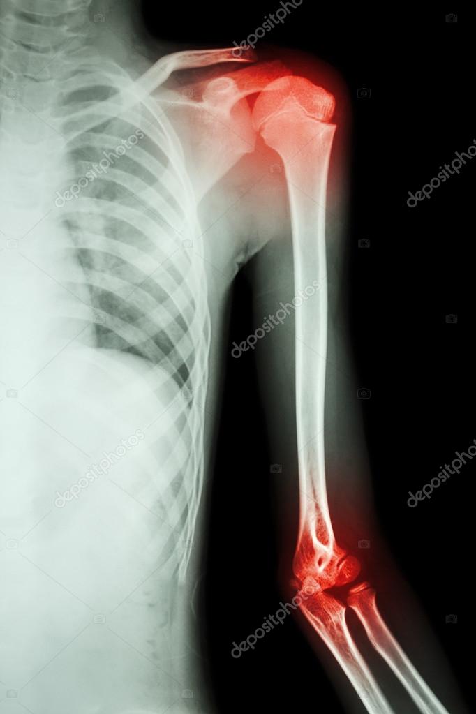
A plain x ray film of the shoulder may show dislocation osteoarthritis or a fracture of the humerus. Quizzes about radiology anatomy quiz.
 Ap Shoulder External Rotation
Shoulder dislocation is a term often used loosely to indicate dislocation of the head of the humerus from the glenoid of the scapula.
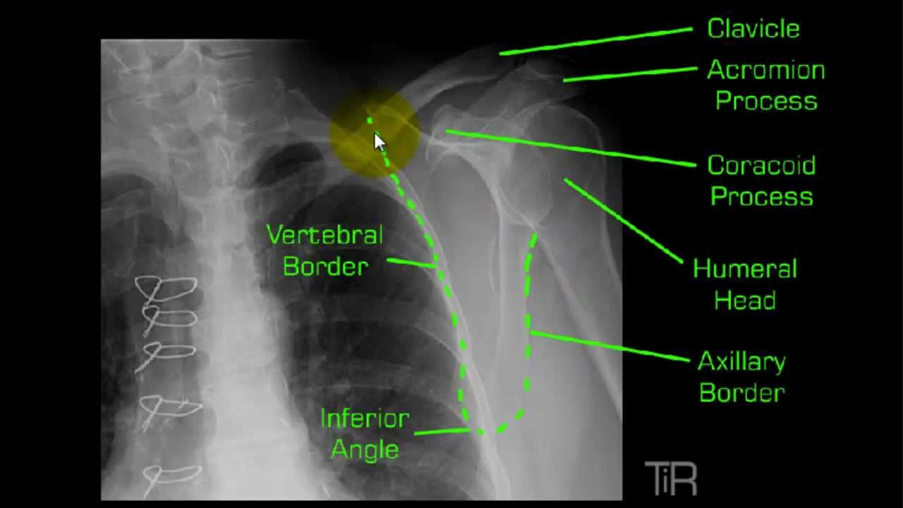
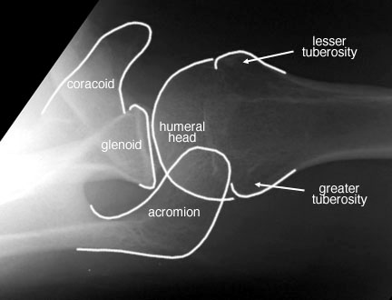
Shoulder x ray anatomy. Drag here to reorder. Annotated anatomy of a lateral shoulder. Anterior dislocations are usually associated with trauma with the arm abducted and in external rotation.
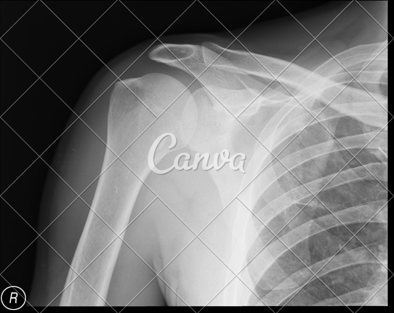
Click here to load quiz. The projection demonstrates the shoulder in its natural anatomical position allowing for adequate radiographic examination of the entire clavicle and scapula as well as the glenohumeral acromioclavicular and sternoclavicular joints of the shoulder girdle. Is to feel for the medial border of the scapula and line it up with the anterior portion of the acromion and x ray straight down the line.
Systematic review choosing a search strategy and utilizing it consistently is a helpful method to overcome common err. Detailed anatomy description of shoulder. Mr arthrography of the shoulder.
5 neck of. Shoulder radiographs are common films to see in the emergency department especially during the weekend after sporting events. Ap internal rotation.
Shoulder ap external rotation. Normal radiographic anatomy of the shoulder. X ray shoulder x ray pelvis x ray hand pa ct head.
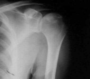
X ray films cannot diagnose muscle or tendon injuries. Standard orthogonal planes can be obtained as a standard ap shoulder radiograph which is taken in external rotation and a lateral view of the scapula. Diagnosis not applicable diagnosis not applicable.
Mri of the shoulder. Shoulder lateral scapula view. Scroll or drag your finger down to reveal the radiographic anatomy for each shoulder view.
Pertinent anatomy how to tell over or under rotation and how to correct. The anatomy of the shoulder and the position required to acquire the lateral film means that modification need to be made for trauma series. Case contributed by dr matt skalski.
This webpage presents the anatomical structures found on shoulder x ray. The shoulder ap view is a standard projection that makes up the two view shoulder series. Shoulder x ray ap projection.
Annotated anatomy of a lateral shoulder figure 4. The shoulder can dislocate posteriorly but anterior dislocation is approximately 50 times more common. Opening the quiz in incognito mode will prevent answers becoming pop up suggestions for future attempts.
 The Shoulder
 Medical Imaging Technology Radiographic Anatomy Of Shoulder

 Diagram Apical Ap Axial Shoulder Anatomy Xray 2018
 Shoulder X Ray Labeled Anatomy Radiology Case


 Shoulder Lateral Canine X Ray Positioning Guide Imv Imaging

Plain Film X Ray Principles Interpretation Teachmeanatomy
 X Schouder Startradiology
 The Shoulder
 Y View Shoulder Mp4
 Normal Shoulder Radiology Case Radiopaedia Org

 Ap Of The Shoulder Radiology Student Shoulder Anatomy
 Shoulder Osteonecrosis Practice Essentials Relevant

 Radiological Anatomy Of The Shoulder Arm Elbow Forearm
 Orthopedic Radiology Dr W Pacheco 2 Xi Ppt Video Online
 Posterior Shoulder Dislocation Litfl Medical Blog Trauma
 Radiographic Anatomy Of Paediatric Shoulder Radiology Imaging

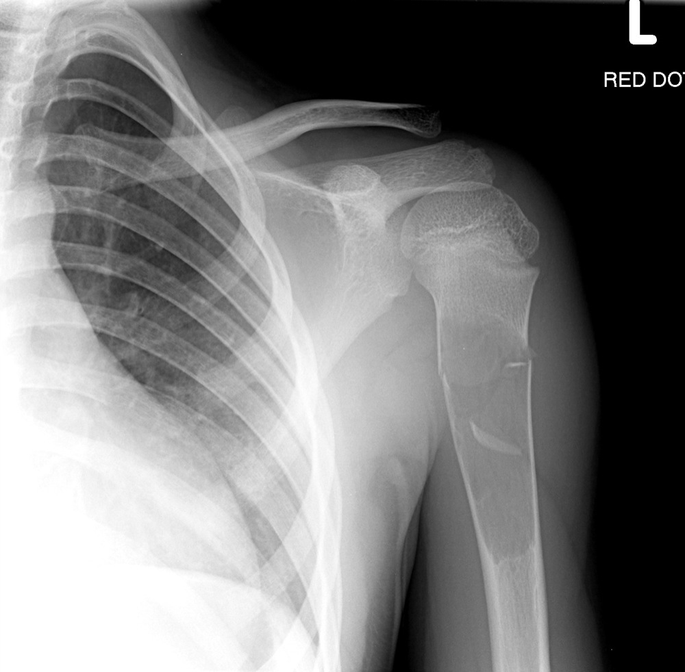
 Left Shoulder X Ray Anatomy Film X Ray Left Shoulder And
 Shoulder Dislocation Core Em
 Ecr 2016 C 1653 Traumatic Shoulder Injuries What
 Anatomy Quiz Shoulder Axial View Radiology Case
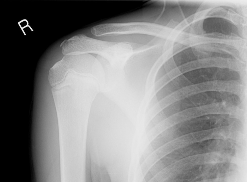
 Right Shoulder X Ray Anteroposterior No Pathologic Changes
Belum ada Komentar untuk "Shoulder X Ray Anatomy"
Posting Komentar