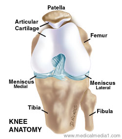
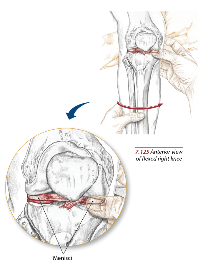
Anatomy Of The Meniscus
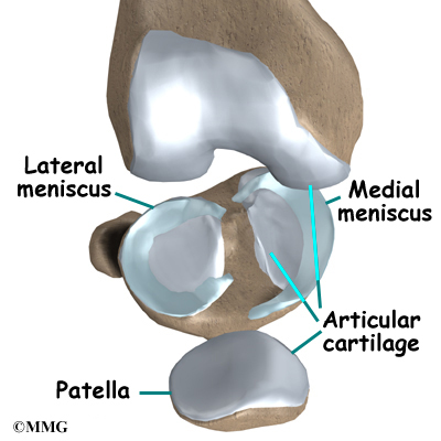
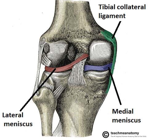
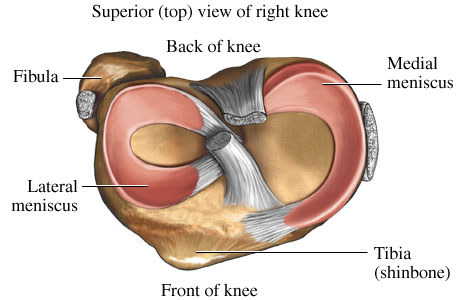
The medial meniscus is the central band of cartilage attached to the tibia or shinbone. The band goes around the knee joint in a crescent shaped path and is located between the medial condyles of the shin and the femur or thighbone.
 Lateral Meniscus An Overview Sciencedirect Topics
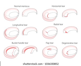
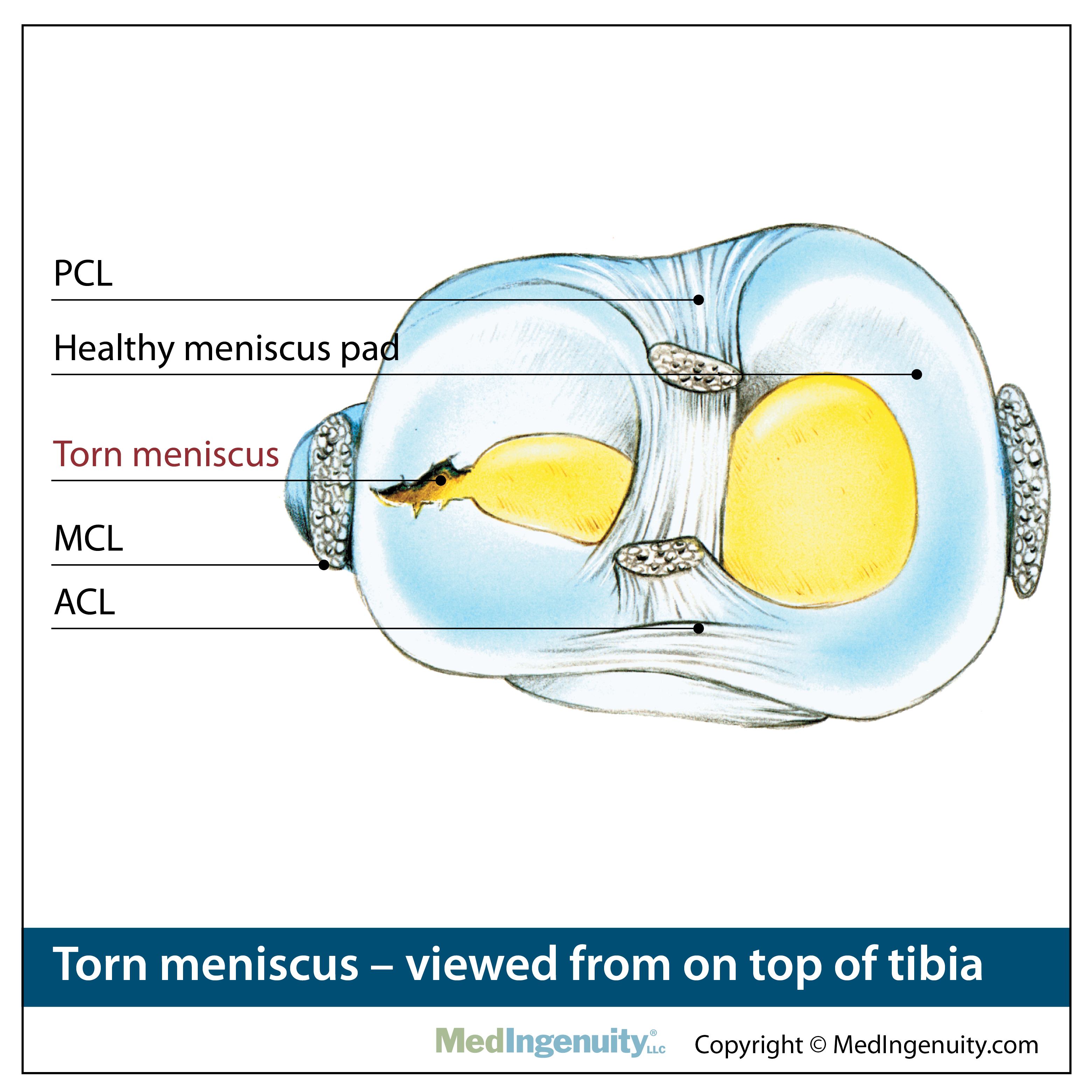
Most tears occur in the back portion posterior horn of the meniscus.

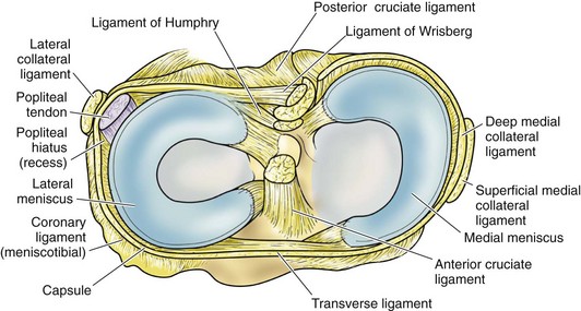
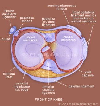
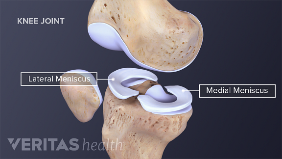
Anatomy of the meniscus. The medial condyles are areas of these bones located on the inner sides of the knees. They are concave on the top and flat on the bottom articulating with the tibia. The lateral meniscus located on the outer side of the knee is shaped like a u.
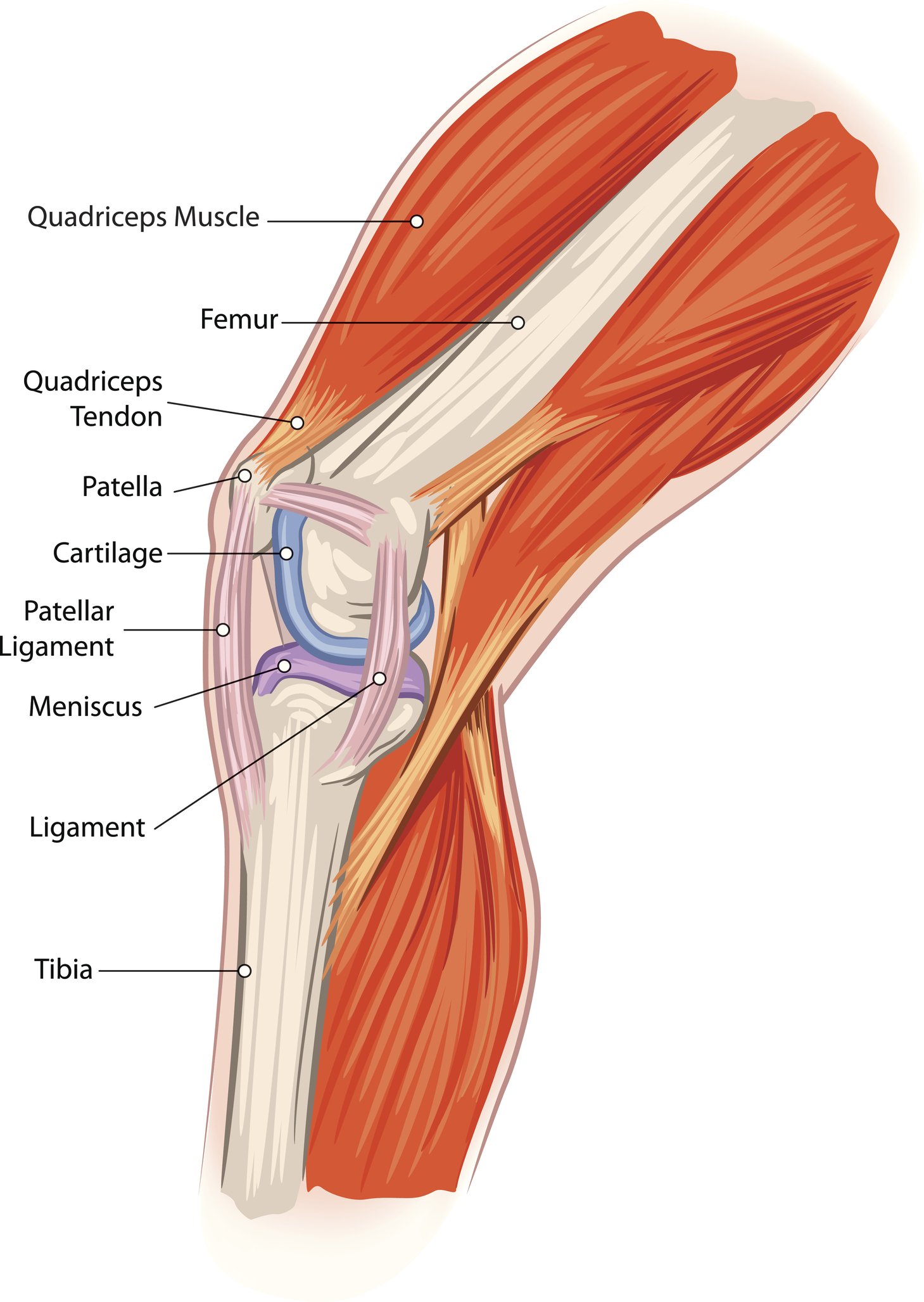
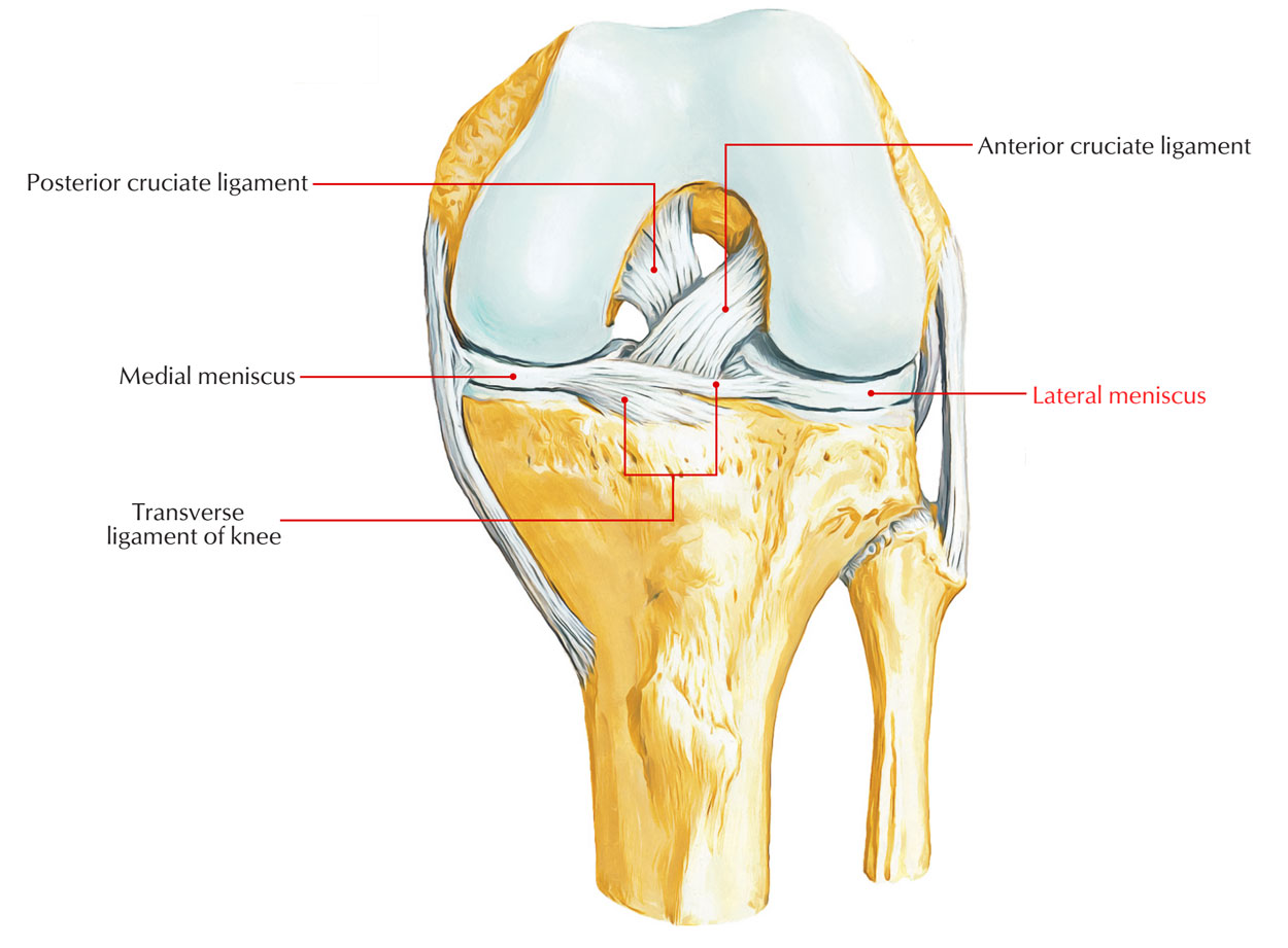
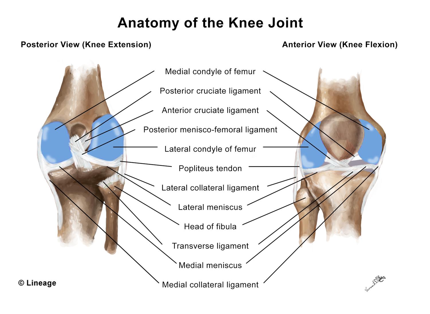
The meniscus is a cushion structure made of cartilage which fits within the knee joint between the tibia and femur. The knee joint contains the meniscus structure comprised of both a medial and a lateral component situated between the corresponding femoral condyle and tibial plateau figure 1. Each is a glossy white complex tissue comprised of cells specialized extracellular matrix ecm molecules and region specific innervation and vascularization.
Is found on the inner side of the knee and is the larger of the two. Some will occur in additional or adjacent locations. The medial meniscus which is located on the inside of the knee is shaped like a c.
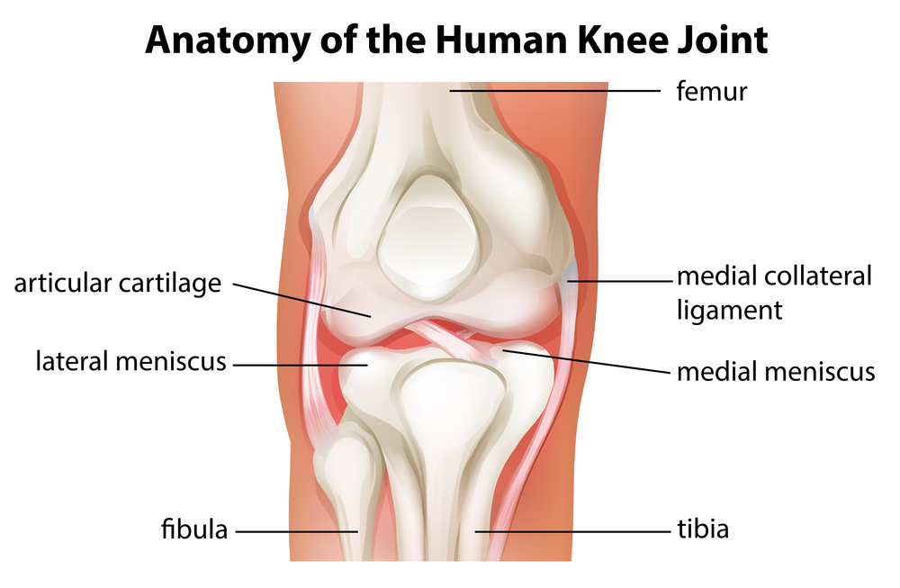
Meniscus anatomy the menisci of the knee are two pads of fibrocartilaginous tissue which serve to disperse friction in the knee joint between the lower leg tibia and the thigh femur. Torn meniscus anatomy. These loose pieces are responsible for the subsequent symptoms.
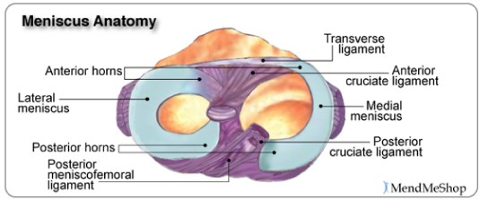
They are attached to the small depressions fossae. Ebraheims educational animated video describes the anatomy of the meniscus. Structure of meniscus two ends both attached to the tibia.
Two surfaces the upper surface is concave for articulation with the femur. Is found on the outer side of the knee. A torn meniscus is just that it is a tear in the structure of the meniscus usually with a loose fragment or several loose pieces.
Each individual meniscus is formed to fit the area of the joint surrounding it. Two borders outer border is thick convex and fixed to the fibrous capsule inner border is thin. The structure of the meniscus 1 medial meniscus.
 Physical Therapy To Treat Torn Meniscus Comparable To
 What Is A Meniscus
 Knee Human Anatomy Function Parts Conditions Treatments
 Can Stem Cells Treat An Acl Tear Or Torn Meniscus
 Easy Notes On Lateral Meniscus Learn In Just 4 Minutes
Adolescent Sports Injuries Of The Knee Cleveland Clinic
 Anatomy Musculoskeletal Key
 Knee Anatomy Including Ligaments Cartilage And Meniscus
 Menisci Of The Knee Joint Healthlink Bc
 Atro Medical Meniscus Vervanging Replacement Atro Medical
 Pin On Yoga
 Lateral Meniscus Wikipedia
 Meniscus Anatomy Wikipedia
Torn Meniscus Symptoms Treatment Mri Test Recovery Time
 Meniscus Knee Sports Orthobullets
 Lateral Meniscus Physiopedia
 The Knee Joint Articulations Movements Injuries
 Menicus Injuries United States The Orthopedic Center
 Anatomy Of The Knee Joint Paley Orthopedic Spine Institute
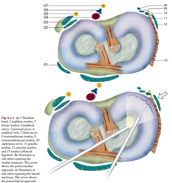 Meniscal Repair Physiopedia
 A Anatomy Of The Meniscus Viewed From Above Adapted Image
 1000 Meniscus Stock Images Photos Vectors Shutterstock
 Meniscus Tear Orthopedics Medbullets Step 2 3
 Meniscus Anatomy And Injuries
 Understanding Meniscus Tears

 Northwest Hills Surgical Hospital Pain From Meniscus Tears
Belum ada Komentar untuk "Anatomy Of The Meniscus"
Posting Komentar