Iris Anatomy
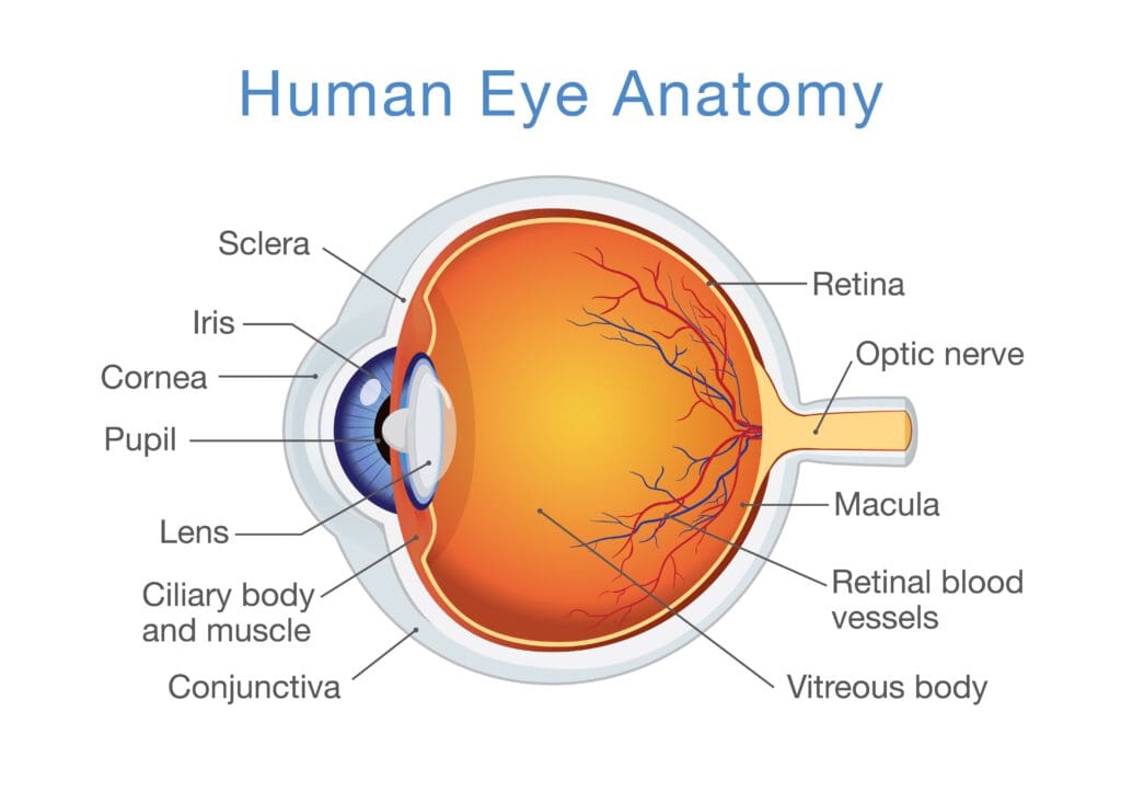
Iris in anatomy the pigmented muscular curtain near the front of the eye between the cornea and the lens that is perforated by an opening called the pupil. This layer extends all the way down to the iris root.
 Iris Anatomy Wikipedia
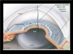
Anatomy and physiology of the iris the iris is a protected internal organ of the eye located behind the cornea and the aqueous humour but in front of the lens.
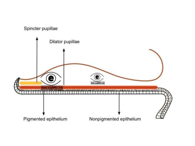
Iris anatomy. In the blue iris the anterior border layer is thin with only a few pigment cells in the brown iris it is thick and densely pigmented. Contains fibroblasts and melanocytes. Choose from 172 different sets of iris anatomy flashcards on quizlet.
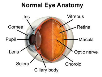
Learn iris anatomy with free interactive flashcards. The iris divides the space in front of the lens into anterior chamber and posterior chamber. It is bathed in front and behind by a fluid known as the aqueous humour.
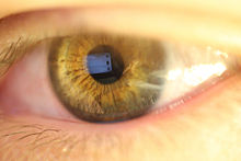
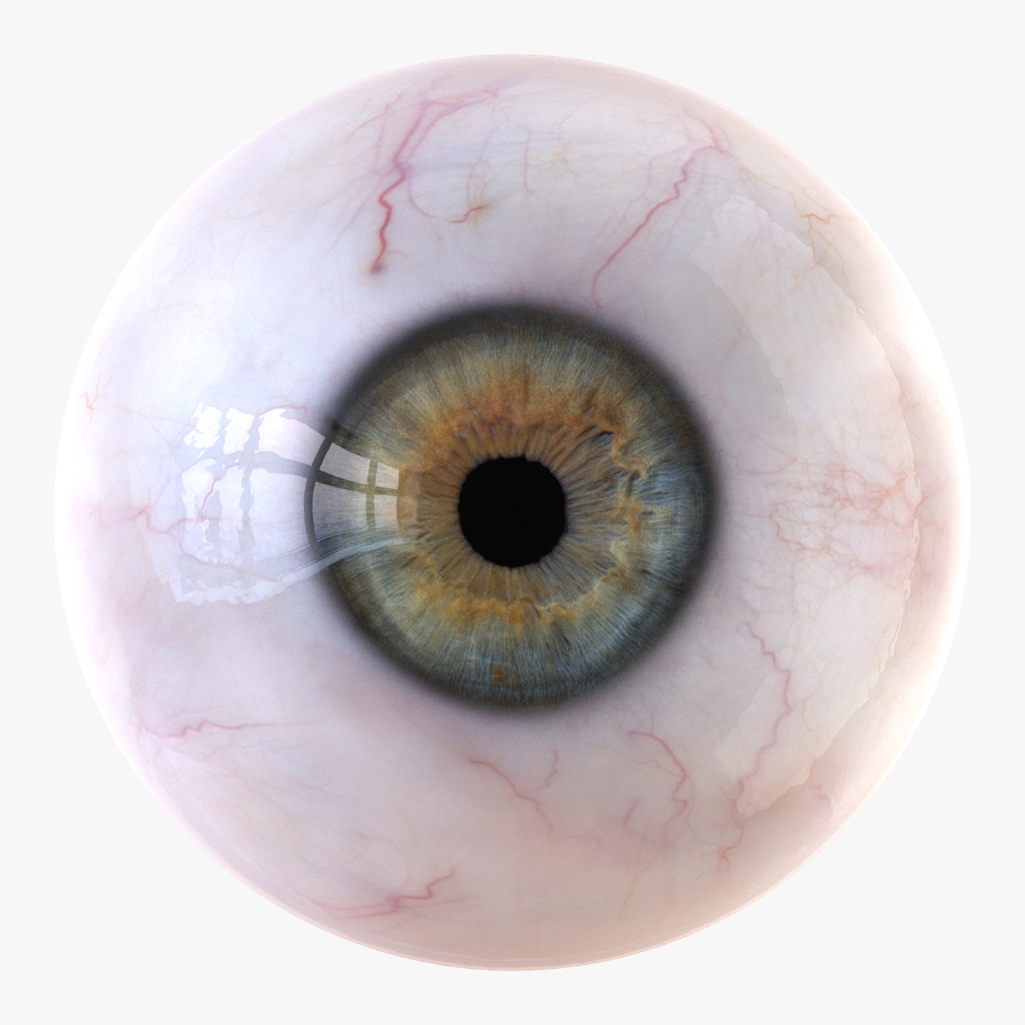
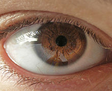
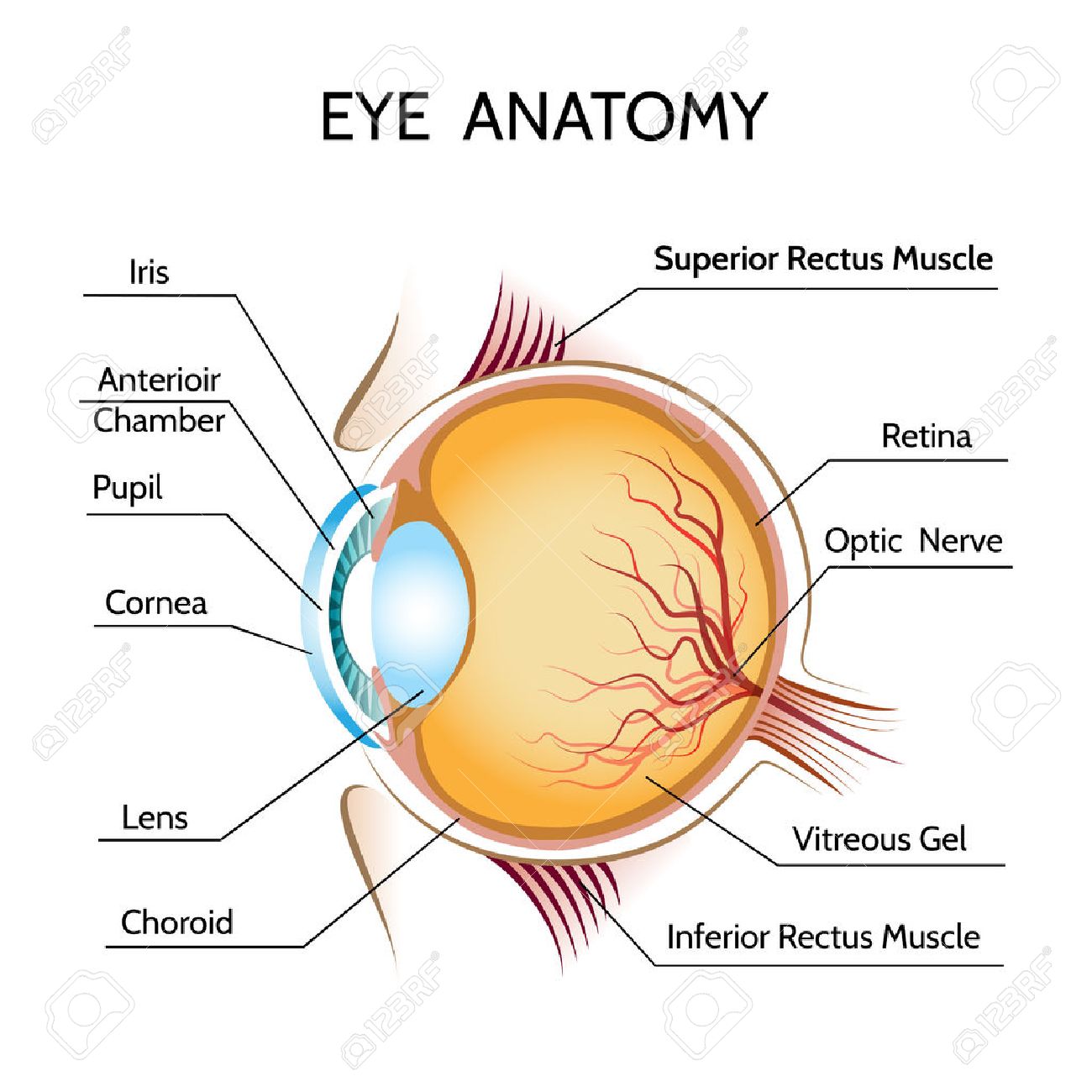
Eye color is the color of the iris which can be green blue or brown. For 18 irides for 23 iris. The iris is located in front of the lens and ciliary body and behind the cornea.
Just behind the iris and pupil lies the lens which helps focus light on the back of your eye. Light projects through your pupil and. Average diameter of the iris is 10 to 11 mm.
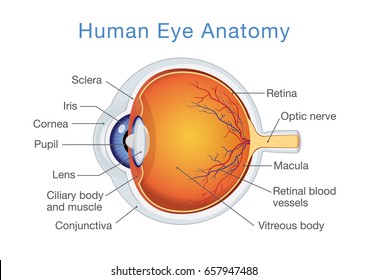
The iris is a flat and ring shaped membrane behind the cornea of the eye with an adjustable circular opening in the center called a pupil. In optical terms the pupil is the eyes aperture while the iris is the diaphragm. It is seen in cross section in the anatomical drawing above.
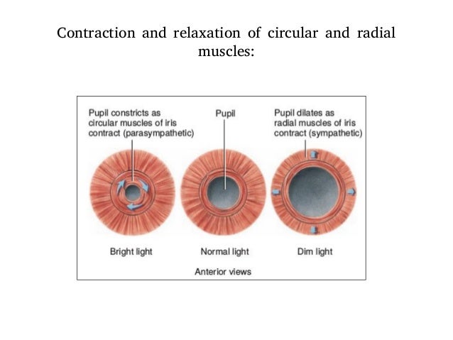
In humans and most mammals and birds the iris is a thin circular structure in the eye responsible for controlling the diameter and size of the pupil and thus the amount of light reaching the retina. A plural of iris. Eye color is defined by that of the iris.
Most of the eye is filled with a clear gel called the vitreous. Iris is attached to the middle of anterior surface of ciliary body. This is the structure that provides an individual with eye.
Iris anatomy synonyms iris anatomy pronunciation iris anatomy translation english dictionary definition of iris anatomy. It is the only internal organ of the body that is normally visible externally. The iris is an anatomical structure in the eye responsible for controlling the diameter and size of the pupils and the amount of light reaching the pupil.
In some cases it can be hazel light brown.
 Anatomy Of The Eye Including Iris Pupil And Cornea
 Iris Anatomy
 Anatomy And Physiology Of Eye

Lasik
.jpg) Eye Anatomy And Eye Diagram Iris Pharma
Eye Anatomy And Eye Diagram Iris Pharma
 Iris Anatomy Wikipedia

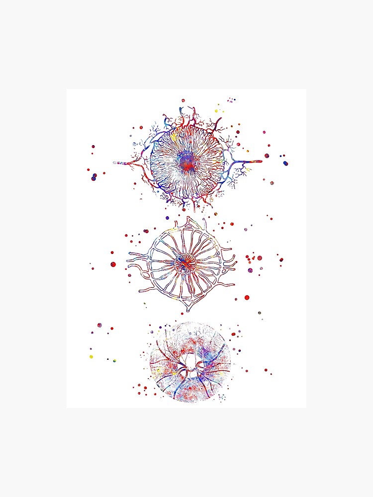
 Human Eye Iris Anatomy Medical Art Watercolor Painting Canvas Art 1796

 Anatomy Of The Eye The Ottawa Hospital
 Eye Anatomy Detail Picture Image On Medicinenet Com
 Anatomy Of The Iris Internal Left And External Right
 Eye Anatomy And Function
 Detailed Human Eye
 Iris Anatomy Wikipedia
![]() Eye Anatomy Muscles Arteries Nerves And Lacrimal Gland
Eye Anatomy Muscles Arteries Nerves And Lacrimal Gland
 Iris Anatomy 2 1 Iris Segmentation 2 1 2 Geodesic Active
![]() Iris Anatomy Wikipedia
Iris Anatomy Wikipedia

 Reflection Of Light Diagram Inspirational Iris Anatomy
 Eye Iris Realistic Anatomy Concept
 Eye Iris Anatomy Biology Backpack By Loissa99
 Anatomy Of Iris
 The Iris Its Anatomy Function Related Eye Diseases

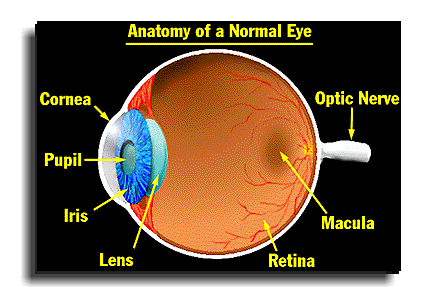 Anatomy Of A Normal Human Eye Amdf
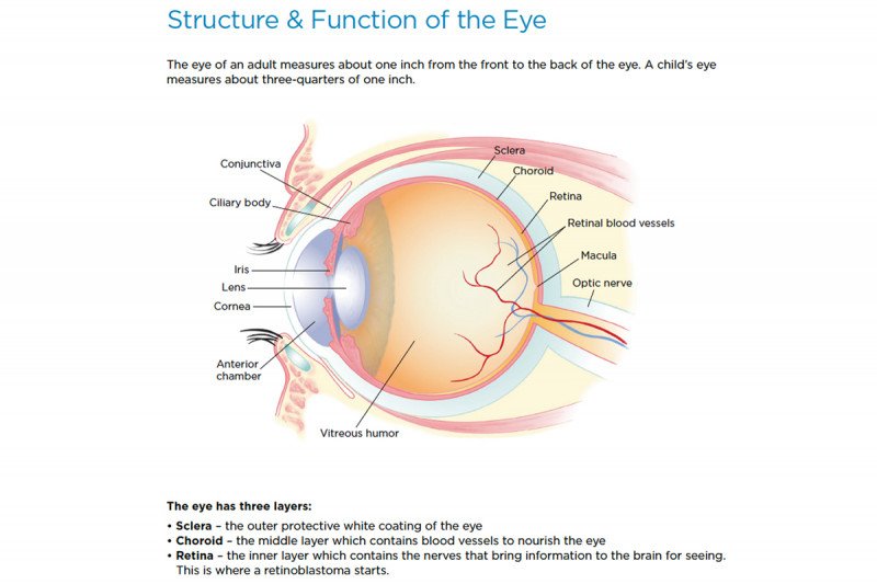 Retinoblastoma Anatomy Of The Eye Memorial Sloan
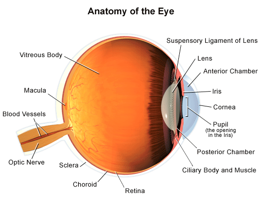 Anatomy Of The Eye
 Human Eye Eye Anatomy Anatomy Of The Iris Blood Supply To Iris Papilla Of The Retina Photographic Print
 Vector Structure Of The Human Eye Anatomy And Medicine Iris
 Imagenes Fotos De Stock Y Vectores Sobre Eye Iris Anatomy
 Eye Anatomy Iris And Optic
 Amazon Com Ambesonne Educational Mouse Pad Human Eye
Belum ada Komentar untuk "Iris Anatomy"
Posting Komentar