Anatomy Of The Knee Bones
The largest joint in the body the knee moves like a hinge allowing you to sit squat walk or jump. They are they soft tissues found at the end of muscles which link the muscle to bone.
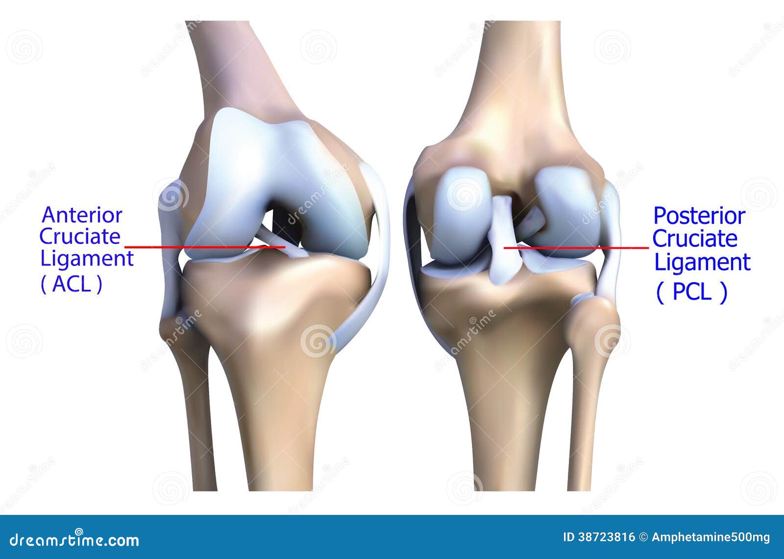 Anatomy Of The Knee Bone And Ligament Stock Illustration
Tendons are often overlooked as part of knee joint anatomy.
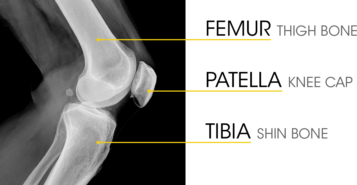
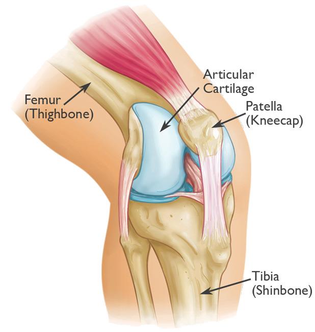
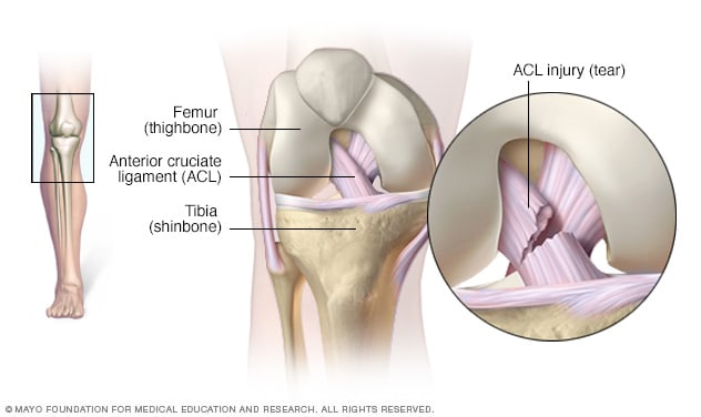
Anatomy of the knee bones. And the patella which is sometimes called the kneecap. The knee is the largest and most complex joint in the body. The femur patella and tibia are all bones within the knee.
The knee is one of the largest and most complex joints in the body. The bones give strength stability and flexibility in the knee. Tibia the bone at the front of the lower leg or shin bone.
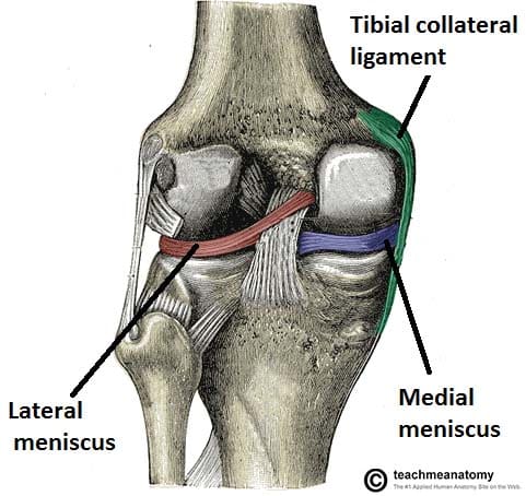
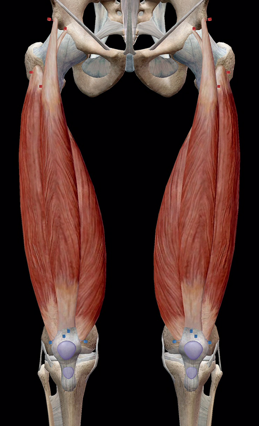
The knee consists of three bones. Participation in sports and recreational activities are risk factors for knee injury. Flexors include the biceps femoris the plantaris the semitendinosus and the semimembranosus.
The smaller bone that runs alongside the tibia fibula and the kneecap patella are the other bones that make the knee joint. Fast facts on knee anatomy. Extensors include the articularis genus the rectus femoris and the quadriceps femoris.
Tibia commonly called the shin bone runs from the knee to the ankle. When there is damage to one of the structures that surrounds the knee joint this can lead to discomfort and disability. The bones of the knee and the leg include the femur which is the large thigh bone.
The top of the tibia is made of two plateaus and a knuckle like protuberance called the tibial tubercle. The knee cap actually sits inside the patellar tendon. Commonly referred to as the kneecap this nearly heart shaped bone at the center of the knee helps extend the knee and protect the joint from impact.
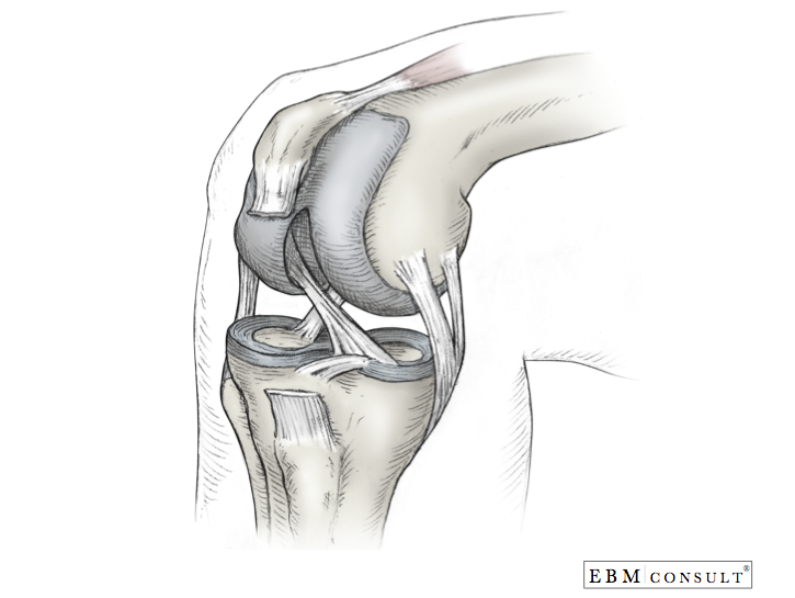
The main tendon found at the knee is the patellar tendon which links the quads muscles to the shin bone. The knee joint is a complex structure that involves bones tendons ligaments muscles and other structures for normal function. Picture of the knee.
The knee is the joint where the bones of the lower and upper legs meet. The tibia and fibula which are the leg bones between the knee and ankle. This long bone runs between the hip and the knee.
The fourth bone of the knee is the patella. Meanwhile the muscles found within the knee include a variety of flexors and extensors. The knee is a synovial joint meaning it contains a fluid filled capsule.
Femur the upper leg bone or thigh bone. The knee joins together the thigh bone shin bone fibula on the outer side of the shin and kneecap. Four bones make up the knee see above image.
The knee joins the thigh bone femur to the shin bone tibia. Bones of the knee.
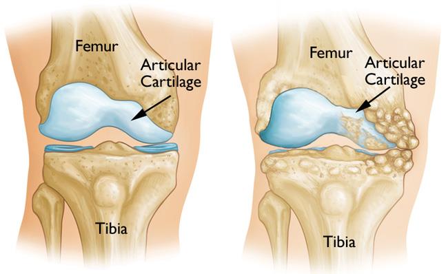
 Understanding The Role Of Cartilage In The Knee
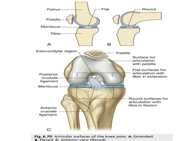 Anatomy And Clinical Importance Of Knee Joint
 Nerves Of The Knee Joint
 Iii Syndesmology 7b The Knee Joint Gray Henry 1918
 Patellar Fractures Broken Kneecap Orthoinfo Aaos
 Anatomy Knee Joint Cross Section Showing The Major Parts Which
 What Are The Parts Of The Knee Joint Systems4knees
Knee Anatomy
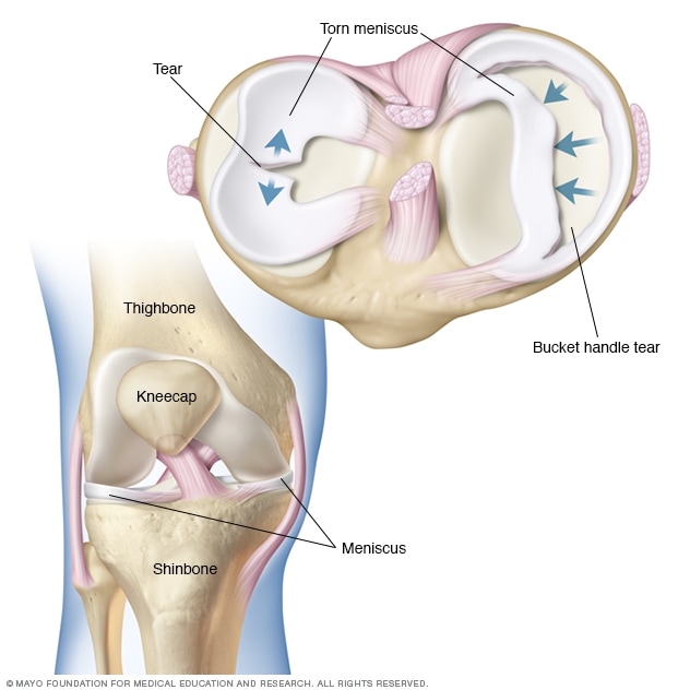 Knee Pain Symptoms And Causes Mayo Clinic
 Knee Joint Picture Image On Medicinenet Com
 Anatomy Of The Knee Comprehensive Orthopaedics
 Anatomy Of The Knee A Simple Understanding Osteopathy
 Anatomy Of The Knee Baxter Regional Medical Center
 Knee Joint Anatomy Pictures And Information
 The Knee Joint Articulations Movements Injuries
 Anatomy Knee
 The Human Knee Joint S Anatomy With Visible Cruciate
 Anatomy Of The Knee Ct Arthrography
 Learn Muscle Anatomy Knee Joint Group
 Knee Pain Symptoms And Causes Mayo Clinic
 Anatomy Of Human Knee Joint
 Articular Capsule Of The Knee Joint Wikipedia
Osteotomy Of The Knee Orthoinfo Aaos
Belum ada Komentar untuk "Anatomy Of The Knee Bones"
Posting Komentar